The Incidence and Risk Factors of Lens-iris Diaphragm Retropulsion Syndrome during Phacoemulsification
Article information
Abstract
Purpose
In the present study, the incidence and risk factors of lens-iris diaphragm retropulsion syndrome (LIDRS) were evaluated.
Methods
Patients who underwent cataract surgery using phacoemulsification between June 2014 and December 2014 were included in the study. The preoperative ocular biometric and intraoperative surgical parameters were examined. The incidence of LIDRS and various risk factors were analyzed using an independent t-test, Pearson's chi-square test, and univariable and multivariable logistic regression analyses.
Results
Among 124 eyes of 124 patients, 100 (80.6%) had no LIDRS and 24 (19.4%) had LIDRS. LIDRS occurred in 13 of 31 vitrectomized eyes (41.9%) and 11 of 93 non-vitrectomized eyes (11.8%). Based on univariable analysis, age (odds ratio [OR], 0.920; p = 0.001), vitrectomized eye (OR, 5.038; p = 0.001), spherical equivalent (OR, 0.778; p < 0.001), axial length (OR, 1.716; p < 0.001), anterior chamber depth (OR, 3.328; p = 0.037), and 3.0 mm vs. 2.2 mm incision size (OR, 4.964; p = 0.001) were statistically significant risk factors associated with the development of LIDRS. Conditional multivariable logistic regression showed that vitrectomized eye (OR, 3.865; 95% confidence interval [CI], 1.201 to 12.436; p = 0.023), long axial length (OR, 1.709; 95% CI, 1.264 to 2.310; p = 0.001), and 3.0 vs. 2.2 mm incision size (OR, 3.571; 95% CI, 1.120 to 11.393; p = 0.031) were significant independent risk factors associated with LIDRS.
Conclusions
LIDRS is a relatively common occurrence and was found to be associated with vitrectomized eye, long axial length, and larger incision size. Evaluating risk factors prior to cataract surgery can help reduce associated morbidity.
Iris stability is an important factor for successful cataract surgery. Poor preoperative or intraoperative mydriasis or damaged iris during surgery may induce pseudophakic macular edema, which can lead to poor outcomes following cataract surgery [1]. Several previous reports on intraoperative floppy-iris syndrome have indicated that this condition can affect the results of cataract surgery and can be associated with systemic sympathetic alpha-1A antagonists such as tamsulosin [234].
In 1992, Zauberman [5] first described a phenomenon that occurs during phacoemulsification and that is characterized by excessive anterior chamber deepening, retropulsion of the iris, and extreme widening of the pupil. In 1994, Wilbrandt and Wilbrandt [6] designated this phenomenon as lens-iris diaphragm retropulsion syndrome (LIDRS) and described its mechanism. Posterior movement of the lens-iris diaphragm causes significant discomfort and pain under topical or intracameral anesthesia, and an excessively deep anterior chamber renders phacoemulsification more difficult for the operating surgeon.
Wilbrandt and Wilbrandt [6] and Cionni et al. [7] suggested possible etiologies of LIDRS such as myopic eyes and reverse pupillary block. Management recommendations for LIDRS have also been suggested in several studies [7891011]. Ghosh et al. [12] reported the incidence and association of LIDRS in patients who underwent vitrectomy. However, no prior reports have discussed the prevalence or risk factors of LIDRS in the general population. Therefore, we determined the prevalence and attributing factors for LIDRS and evaluated the efficacy of a bottle-lowering procedure for the management of LIDRS.
Materials and Methods
In this retrospective comparative study, the incidence and risk factors of LIDRS were evaluated in patients who underwent phacoemulsification cataract surgery. The patients received information regarding the procedure and provided informed consent. The study was performed at Samsung Medical Center, Seoul, Korea, in accordance with the Declaration of Helsinki and was approved by the iInstitutional review board of Samsung Medical Center.
The medical records of patients having unilateral cataract and who visited the outpatient department at Samsung Medical Center between June 2014 and December 2014, were retrospectively reviewed. Patients were excluded if they had a history of iris surgery or iris pathology such as iridocyclitis or iris neovascularization or other ocular procedures other than vitrectomy such as trabeculectomy, strabismus surgery, scleral buckling, or intravitreal injection. Exclusion criteria also included traumatic cataract, zonulysis, or any condition that would obstruct the exact evaluation of LIDRS, such as general or retrobulbar anesthesia.
All patients underwent routine phacoemulsification cataract surgery without complications; all surgeries were performed performed by the same skilled surgeon (ESC).
Preoperative ocular biometric parameters
Before the surgery, patients underwent full ophthalmological examinations including uncorrected distance visual acuity, corrected distance visual acuity, manifest and cycloplegic refractions, slit lamp evaluation, tonometry, gonioscopy, keratometry, corneal pachymetry and topography (Orbscan II; Bausch & Lomb, Rochester, NY, USA), noncontact specular microscopy (SP-8000; Konan Medical, Nishinomiya, Japan), and binocular indirect ophthalmoscopy with the pupils dilated. Information regarding reoperative risk factors was obtained and evaluated, including sex, age, history of diabetes or hypertension, history of vitrectomy, spherical equivalent, and axial length and anterior chamber depth (defined as corneal epithelium to anterior crystalline lens surface) measured using IOLMaster (Carl Zeiss Meditec, Jena, Germany).
Surgical procedures and intraoperative surgical parameters
A standard dilating regimen consisting of three rounds of 2.5% topical phenylephrine and 1% tropicamide (Mydrin-P; Santen Pharmaceuticals, Osaka, Japan) followed by instillation of proparacaine HCl 0.5% (Alcaine; Alcon Laboratories, Fort Worth, TX, USA) every 5 minutes was used. In all cases, cataract surgeries were performed using a 2.2- or 3.0-mm-sized clear corneal incision, continuous curvilinear capsulorrhexis, phacoemulsification using the Infiniti system (Alcon Laboratories), cortex removal using a bimanual irrigation/aspiration tip, and posterior chamber foldable intraocular lens implantation. Viscoelastics were used to maintain the anterior chamber and mechanically hold the pupil open.
Additional intraoperative factors including incision site (superior vs. temporal), incision size (3.0 mm vs. 2.2 mm), and ophthalmic viscosurgical device (Healon 5; Advanced Medical Optics, Santa Ana, CA, USA vs. Discovisc, Alcon Laboratories) were investigated.
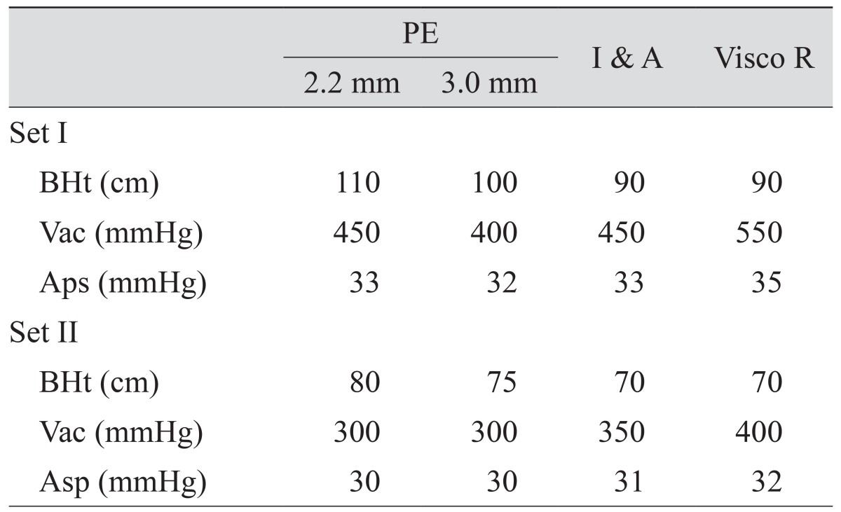
The presence of LIDRS was evaluated three times, during the phacoemulsification, bimanual irrigation and aspiration of residual cortex, and bimanual irrigation and aspiration of ophthalmic viscosurgical devices. LIDRS was graded on a scale from 0 to 3: grade 0, no LIDRS; grade 1, mild pupil dilatation; grade 2, pupil dilatation and patient discomfort; grade 3, abrupt pupil dilatation and patient pain. When LIDRS occurred, the bottle height, vacuum pressure, and aspiration pressure were reduced from set I to set II, and the effect on LIDRS was recorded (Table 1).
Statistical analysis
The data analysis was performed using PASW ver. 18.0 (SPSS Inc., Chicago, IL, USA). The absolute frequency (n) and relative frequency (%) were computed for qualitative variables, and the mean and standard deviations were computed for quantitative variables. The clinical characteristics of patients with and without LIDRS were compared using Pearson's chi-square test for categorical variables and the independent t-test for continuous parameters.
The 95% confidence intervals (CIs) for odds ratios (ORs) were calculated. Univariable simple logistic regression analysis was used to examine the associations between LIDRS and the variables. Factors with a p-value <0.2 were considered associated with LIDRS and were included as candidates in multivariable analysis. Stepwise conditional multivariable logistic regression analysis was performed to evaluate meaningful risk factors affecting LIDRS. A p-value <0.05 was considered statistically significant.
Results
This study consisted of 124 eyes of 124 patients, including 80 females and 44 males, with a mean age of 62.5 ± 11.1 years (range, 31 to 92 years). All surgeries were uneventful. Twenty-four eyes (19.4%) developed LIDRS, which were 13 of 31 vitrectomized eyes (41.9%), and 11 of 93 non-vitrectomized eyes (11.8%). Table 2 shows the incidence and most severe grade of LIDRS during the phacoemulsification, irrigation and aspiration of the residual cortex, and residual viscoelastic removal.
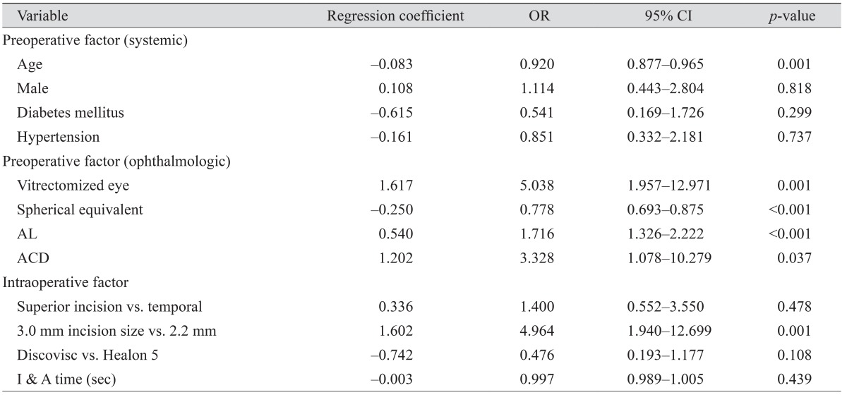
Table 3 shows the associations between LIDRS and clinical characteristics. Table 4 shows the ORs for clinical characteristics associated with LIDRS based on univariable logistic regression analysis. In brief, statistically significant associations with LIDRS were found for age (OR, 0.920; 95% CI, 0.877 to 0.965; p = 0.001), vitrectomized eye (OR, 5.038; 95% CI, 1.957 to 12.971; p = 0.001), spherical equivalent (OR, 0.778; 95% CI, 0.693 to 0.875; p < 0.001), axial length (OR, 1.716; 95% CI, 1.326 to 2.222; p < 0.001), anterior chamber depth (OR, 3.328; 95% CI, 1.078 to 10.279; p = 0.037), and 3.0 mm incision size (vs. 2.2 mm; OR, 4.964; 95% CI, 1.940 to 12.699; p = 0.001). Multivariable data analysis using conditional stepwise logistic regression analysis of the variables showed that vitrectomized eye (OR, 3.865; 95% CI, 1.201 to 12.436; p = 0.023), long axial length (OR, 1.709; 95% CI, 1.264 to 2.310; p = 0.001), and 3.0 mm incision size (vs. 2.2 mm; OR, 3.571; 95% CI, 1.120 to 11.393; p = 0.031) were significant independent factors associated with LIDRS (Table 5).

The results of univariable logistic regression for potential risk factors of LIDRS during phacoemulsification

The result of multivariable logistic regression for significant risk factors of LIDRS during phacoemulsification
Among the 24 eyes that experienced LIDRS, bottle height, vacuum pressure, and aspiration pressure were reduced from set I to set II in 15 eyes, nine eyes (60%) showed a decrease in the grade of LIDRS, and six eyes (40%) showed no interval change.
Discussion
LIDRS is an emerging problematic condition that can increase surgical challenges and patient morbidity. Although this condition has been attributed to an excessively deep anterior chamber, iris retropulsion, and extreme widening of the pupil during small incision cataract surgery, the incidence and factors that contribute to this condition remain unclear, and no strategy for management of patients has been clearly defined. LIDRS increases surgical challenges and may be associated with significant discomfort for patients receiving phacoemulsification under topical or intracameral anesthesia [7].
LIDRS is a well-recognized condition in myopic eyes and eyes that undergo vitrectomy [12]. However, previous clinical reports have not discussed the presentation of LIDRS, and the incidence and risk factors of LIDRS in the non-vitrectomized general population remain unknown. In the present study, the incidence of LIDRS and associated risk factors were evaluated.
The incidence of LIDRS in eyes that previously underwent vitrectomy has been reported in a few studies, with rates ranging from 0% to 100% [12131415161718]. In our study, among 31 vitrectomized eyes, 13 (41.9%) experienced some degree of LIDRS during phacoemulsification surgery. Bardoloi et al. [14] evaluated the occurrence of LIDRS in myopic patients and found that it occurred in eight of 52 non-vitrectomized myopic eyes (15.4%). The present study is the first report on the incidence of LIDRS in the non-vitrectomized general population of patients that underwent phacoemulsification cataract surgery; we observed an incidence of 11.8%.
Only patients of Korean ethnicity were enrolled in this study. Most Koreans have a dark brown iris with light brown eyes; individuals without a brown iris are extremely rare. Studies have indicated that iris color may contribute to iris stability; hence, the incidence of LIDRS can vary based on genetic differences in iris color [19].
Predicting LIDRS before phacoemulsification cataract surgery is important for both cataract surgeons and patients. Univariable analysis showed that the variables significantly correlated with or associated with LIDRS (p < 0.2) were age, vitrectomized eye, spherical equivalent, axial length, anterior chamber depth, 3.0 mm incision size (vs. 2.2 mm), and use of Discovisc as an ophthalmic viscosurgical device (vs. Healon 5). Among these, the best fit conditional multiple logistic regression model showed that vitrectomized eye, long axial length, and 3.0 mm incision size (vs. 2.2 mm) were statistically significant at a 0.05 significance level.
Previous vitrectomy and axial myopia are well-known risk factors of LIDRS [121920]. Ghosh et al. [12] reported the LIDRS severity was significantly greater in extensive vitrectomy than in limited vitrectomy (adjusted OR, 4.42; 95% CI, 1.19 to 16.45; p = 0.027) and with increasing axial length (adjusted OR, 3.05; 95% CI, 1.86 to 5.01; p < 0.001). In our study, the significance of both risk factors were demonstrated using multivariable logistic regression analysis, with ORs of 3.865 (95% CI, 1.201 to 12.436; p = 0.023) in the vitrectomized eye and 1.709 (95% CI, 1.264 to 2.310; p = 0.001) in the long axial length. Ghosh et al. [12] proposed that low-viscosity vitreous fluid that leaked into the anterior chamber during the initial stage of surgery in patients with axial myopia, vitreous degeneration, and a history of vitrectomy resulted in a relative volume deficit in the vitreous cavity.
Our multivariate analysis indicated that the incidence of LIDRS increased in eyes with previously known risk factors such as previous vitrectomy and long axial length. In addition, new associations were identified, including a relatively large corneal incision width of 3.0 mm compared with 2.2 mm. The direction and size of the clear corneal incision were determined based on the axis and amount of corneal astigmatism. We measured corneal astigmatism using manual keratometry, automated keratometry with optical biometry (IOLMaster), and corneal topography; all three methods gave comparable results. An incision was made on either the temporal or superior side, depending on which had a steeper axis. If the corneal astigmatism was greater than 0.75 D, a 3.0 mm incision was made with a 3.0 mm angled diamond keratome. If the corneal astigmatism was equal to or less than 0.75 D, a 2.2 mm incision was made with a 2.2 mm angled diamond keratome.
Two possible hypotheses can explain the increase in LIDRS with a relatively larger clear corneal incision. In the present study, most LIDRS during phacoemulsification occurred during the initial phase, especially after insertion of the phacoemulsification tip. A greater phacoemulsification sleeve volume through a larger incision may cause relative overload of the volume and pressure in the anterior chamber compared to the vitreous cavity. The differences may also be explained by iris stability depending on incision size. Corneal incisions are characterized by three parameters: construction, location, and size. All of the surgeries were performed by the same surgeon using the same technique; therefore, significant differences in clear corneal incision wound construction were unlikely. Hence, wound stability may be associated with wound size. Smaller wounds reduce iris prolapse, allow less leakage during surgery, and increase chamber stability. Therefore, the relative instability of a larger wound may explain the occurrence of LIDRS during the middle phacoemulsification and irrigation/aspiration stages.
Several guidelines have been suggested for managing LIDRS based on the characterization by Wilbrandt and Wilbrandt [6]. Cionni et al. [7] reported that mechanical separation of the iris and capsule is helpful for managing reverse pupillary block, an essential mechanism of LIDRS. Nahra Saad et al. [8], Lee et al. [9], and Vishwanath [10] used an iris hook retractor to lift the iris and relieve the pupillary block. Packard introduced a series of maneuvers, bottle lowering, and a side-port instrument to prevent LIDRS in high-risk patients [11]. Ghosh et al. [12] suggested that lowering of parameters, slight bottle height reduction, and a corresponding reduction in maximum aspiration rate could be applied to avoid anterior chamber collapse in cases at risk of LIDRS. We evaluated the effects of bottle- and pressure-lowering procedures in several patients and observed these techniques were effective in nine of 15 eyes (60%).
This study was limited by the retrospective design and the insufficient evaluation of various management techniques. Although all specific events such as LIDRS or intraoperative floppy iris syndrome during surgery were recorded, there is a possibility that cases were missed. We managed all cases without complications but were unable to assess all possible risk factors or evaluate the effects of bottle- and pressure-lowering procedures in all cases. Additional prospective interventional large-scale studies are needed to verify the application of our proposed management guidelines.
Our study results indicate the incidence of LIDRS is one fifth (20%) in the general population. LIDRS occurred in 41.9% of vitrectomized eyes and 11.5% of non-vitrectomized eyes. The results indicated that young age, history of vitrectomy, myopic spherical equivalent, long axial length, deep anterior chamber depth, and large corneal incision size (3.0 mm vs. 2.2 mm) were associated with LIDRS; specifically, vitrectomized eye, long axial length, and 3.0 mm incision size (vs. 2.2 mm) were determined to be significant independent factors.
Acknowledgements
Involved in conception and design (DHL, ESC, TYC) and conducted the study (DHS, DHL, GH); collection, management, and interpretation of data (DHL, DHS, GH); data analysis (DHL, DHS, GH); writing the manuscript (DHL, DHS); preparation, review, and approval of the manuscript (DHL, ESC, TYC).
Notes
Conflict of Interest: No potential conflict of interest relevant to this article was reported.


