Ultrawide-field Fluorescein Angiography for Evaluation of Diabetic Retinopathy
Article information
Abstract
Purpose
To investigate the advantages of ultrawide-field fluorescein angiography (FA) over the standard fundus examination in the evaluation of diabetic retinopathy (DR).
Methods
Ultrawide-field FAs were obtained in 118 eyes of 59 diabetic patients; 11 eyes with no DR, 71 eyes with nonproliferative diabetic retinopathy (NPDR), and 36 eyes with proliferative diabetic retinopathy (PDR), diagnosed by the standard method. The presence of peripheral abnormal lesions beyond the standard seven fields was examined.
Results
Ultrawide-field FA images demonstrated peripheral microaneurysms in six (54.5%) of 11 eyes with no DR and all eyes with moderate to severe NPDR and PDR. Peripheral retinal neovascularizations were detected in three (4.2%) of 71 eyes with NPDR and in 13 (36.1%) of 36 eyes with PDR. Peripheral vascular nonperfusion and vascular leakage were found in two-thirds of eyes with severe NPDR and PDR.
Conclusions
Ultrawide-field FA demonstrates peripheral lesions beyond standard fields, which can allow early detection and a close evaluation of DR.
In diabetic retinopathy (DR), retinal vasculopathy can occur anywhere in the fundus and is usually present in multiple lesions. The presence of DR, even in its mildest form, is associated with risk of visual loss, and life-threatening systemic vascular complications such as stroke, coronary heart disease, and heart failure. It is an independent cardiovascular risk factor [1].
Seven standard fields images for the Diabetic Retinopathy Study are able to create a montage of around 75 degrees of fundus with the standard fundus camera [2,3] and has been used for diagnosis and classification of diabetic retinopathy. Imaging of the peripheral retina is difficult with standard fundus images because of the narrow angle of the fundus camera and peripheral blur caused by combination of reflections and astigmatism [2].
Fluorescein angiography (FA) is better in detecting retinal vasculopathy than color photography. Moreover, the contact lens assisted wide-angle FA, enables up to 160 degrees field of view [4], although it requires a high technical capability. Optomap fa (Optos plc, Dunfermline, UK) is a confocal laser scanning ophthalmoscope providing ultrawide-field images with an up to 200 degree field of view, by which simultaneous FA of the posterior pole and periphery of the fundus can be possible. It has been reported that it has substantial benefits for the evaluation of peripheral retinal lesions in uveitis, proliferative diabetic retinopathy, and retinal detachment [2,3,5-7].
We investigated the possible advantage of ultrawide-field FA (UWFA) over standard fundus examinations in detection and evaluation of DR.
Materials and Methods
UWFA images were obtained in 118 eyes of 59 patients diagnosed with diabetic mellitus in the Samsung Medical Center. Patients were classified according to the International Clinical Diabetic Retinopathy Disease Severity Scale. Eleven eyes had no retinopathy, 71 eyes had non-proliferative diabetic retinopathy (NPDR; 19 eyes with mild NPDR, seven eyes with moderate NPDR, and 45 eyes with severe NPDR), and 36 eyes had proliferative diabetic retinopathy (PDR). Images of UWFA beyond the seven standard fields were evaluated for abnormal lesions such as peripheral microaneurysms, nonperfusion, vascular leakages, and neovascularization.
Results
In six (54.5%) out of 11 eyes that did not have DR on standard fundus examination, peripheral microaneurysms were found in UWFA (Fig. 1A and 1B). Peripheral microaneurysms were observed in 84.2% (16 / 19) of eyes with mild NPDR, and all eyes with moderate to severe NPDR and PDR.
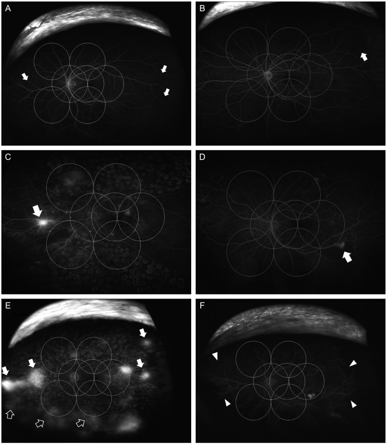
(A,B) Ultrawide-field fluorescein angiography shows peripheral microaneurysms (arrows) outside the seven standard Early Treatment Diabetic Retinopathy Study (ETDRS) fields in eyes diagnosed as normal fundus with the standard fundus examination. (C,D) Peripheral retinal neovascularizations (arrows) were found outside the standard seven ETDRS fields in eyes diagnosed as nonproliferative diabetic retinopathy by the standard method. (E,F) Multiple lesions of neovascularization (arrows), vascular leakage (blank arrows), and nonperfusion (arrow heads) observed beyond the borders of standard fields in ultrawide-field fluorescein angiography.
Peripheral retinal neovascularization was identified beyond the standard fields (Fig. 1C and 1D) in three (4.2%) out of 71 NPDR eyes; none in mild NPDR, one (14.3%) in moderate NPDR, and two (4.4%) in severe NPDR. Peripheral retinal nonperfusion and vascular leakage were found in 47.9% (34 / 71) and 38% (27 / 71) of NPDR eyes, respectively. They were observed only in severe NPDR; nonperfusion in 75.6% (34 / 45), vascular leakage in 60% (27 / 45). Retinal neovascularization, vascular leakage, and nonperfusion in the peripheral retina (Fig. 1E and 1F) were detected in 36.1% (13 / 36), 69.4% (25 / 36), and 63.9% (23 / 36) of PDR eyes, respectively. Table 1 shows peripheral lesions found outside standard fields by UWFA. Peripheral nonperfusion areas were found with sustained macular edema that was refractory to recurrent photocoagulation and intravitreal steroid or anti-VEGF injections in eight out of ten eyes with macular edema without peripheral nonperfusion.
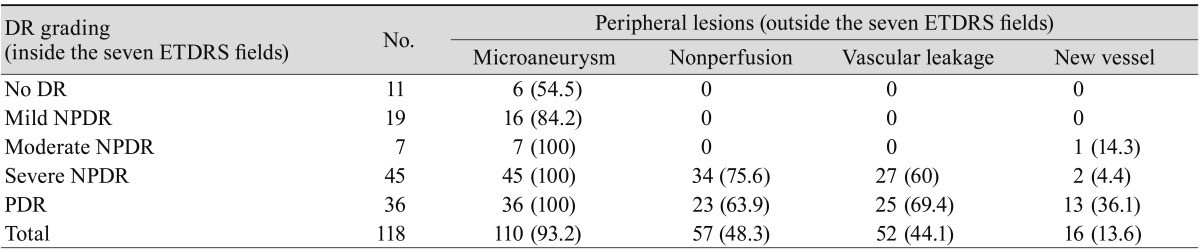
Peripheral lesions beyond the seven ETDRS fields
Discussion
Currently, the standard diagnosis and staging of DR were made according to fundus findings at the posterior retinal area by the fundus examination and fundus photography. Although FA can provide more accurate information of retinal vasculopathy, it has not been used routinely in the examination of diabetic patients due to expense, invasiveness, and the limitation of fundus field. UWFA can provide a wider view than standard FA with better reproducibility, and the current study used UWFA to investigate the usefulness of fluorescein angiography in the diagnosis and classification of DR. UWFA detected peripheral microaneurysms in more than half of diabetic patients diagnosed as no DR in this study. Because a DR lesion is associated with retinal ischemia [4,8], and retinal ischemia is known as the predictive factor of systemic vascular complications [1], it is reasonable to perform UWFA in the subgroup of patients diagnosed as no DR. Patients with a long history of diabetes mellitus and poor control of blood glucose may become ideal candidates. In severe NPDR and PDR patients, UWFA detected new peripheral vessels, and other findings associated with ischemia. However, its clinical significance appears to be questionable in these patients, because the detection rate in the severe NPDR appeared to be quite low, and it may not be helpful to treat eyes with PDR. It is more reasonable to perform UWFA in a certain subgroup of patients, such as patients with chronic, refractory macular edema, and patients with unexplained recurrent vitreous hemorrhage after panretinal photocoagulation. It is known that the extent of nonperfused area anterior to equator is usually wider than the posterior area. The evaluation of ischemia related peripheral retinal findings such as nonperfusion and vascular leakage should be evaluated in patients with chronic, refractive macular edema.
The current study has many limitations, such as small sample size and no control data and is a retrospective cross-sectional study that was not randomized. This study is ultimately a pilot study.
In conclusion, UWFA has significant advantages in detecting and evaluating peripheral abnormal retinal lesions over the standard method because of a far wider angle encompassing almost the entire fundus. More studies for the clinical relevance of fundus peripheral lesions observed through UWFA are needed in the evaluation of the progression, development, diagnosis, and treatment of DR.
Notes
No potential conflict of interest relevant to this article was reported.