Large Thrombosed Basilar Artery Aneurysm Presenting with Homonymous Left Hemianopia: A Case Report
Article information
Dear Editor,
The basilar artery, a major blood vessel in the brain, supplies blood to the brainstem and posterior cranial fossa, especially the cerebellum and occipital lobes. The basilar artery is also responsible for the blood supply of the auditory and vestibular nuclei, as well as the cranial nerve nuclei that control the muscles of the head and neck.
There are two main complications associated with the basilar artery: an occlusion of the artery and an aneurysm. The occlusion of the basilar artery can give rise to serious neurological symptoms such as dizziness, double vision, difficulty speaking, and paralysis or even death if left untreated [1]. The basilar artery aneurysm can result in ocular movement disorders such as sixth nerve palsy, horizontal gaze palsy, skew deviation, and internuclear ophthalmoplegia due to its compression of mid-brain or pons [2]. While diplopia is common in the case of basilar artery aneurysm, visual field defect is very rare, with only few previous reports. To our knowledge, this is the first report of an incidentally discovered thrombosed basilar artery aneurysm with an isolated visual field defect of homonymous left hemianopsia without any other symptoms in Korea.
This study was approved by the Institutional Review Board of Chung-Ang University Gwangmyeong Hospital (2308-102-080) and conducted in accordance with the tenets of the Declaration of Helsinki. Written informed consent for publication of the research details was obtained from the patient.
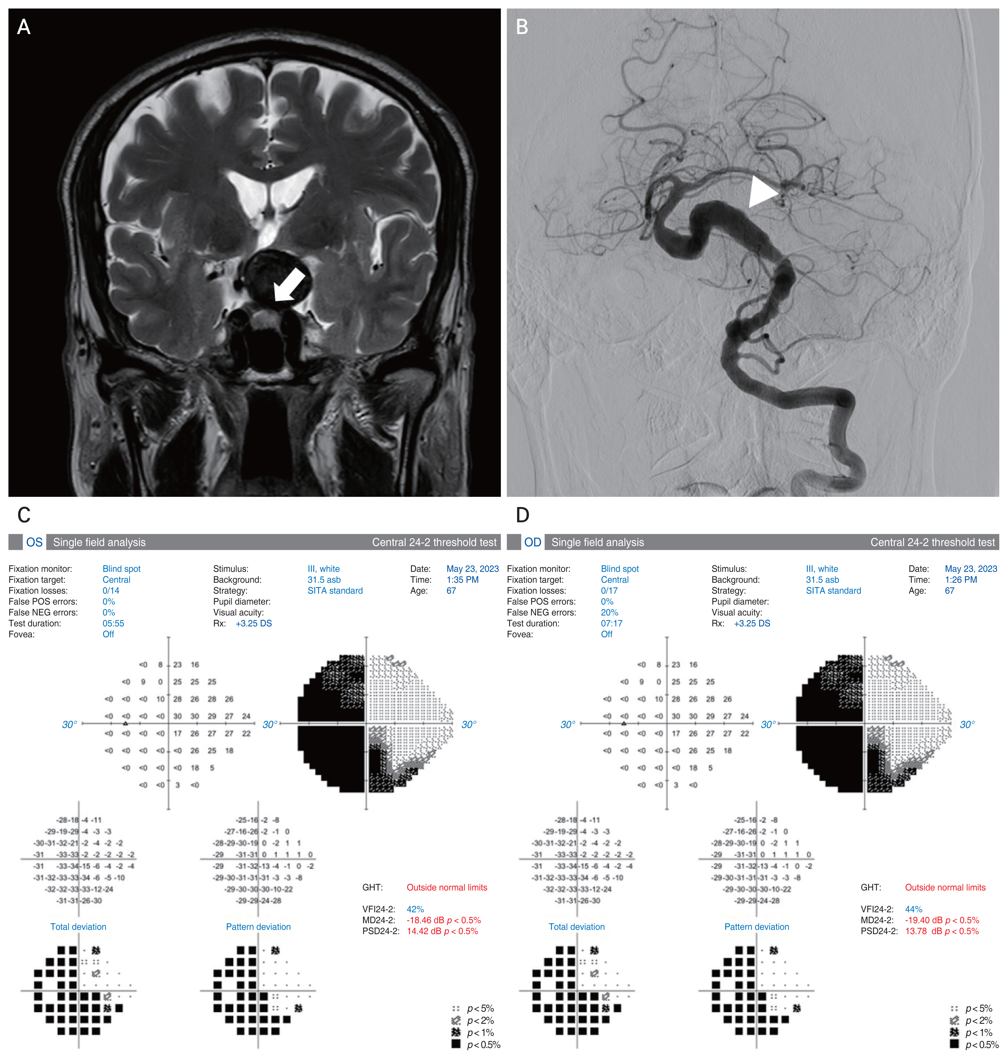
A 67-year-old man visited Chung-Ang University Gwangmyeong Hospital (Gwangmyeong, Korea), complaining of nonspecific visual disturbance fluctuating throughout the day and night for the duration of 6 months. His best-corrected visual acuity was 1.0 in both eyes, and ocular movements were full. There was no abnormal finding in the central nervous system examination, but the brain magnetic resonance imaging (MRI) and four-vessel angiography revealed an unruptured large fusiform basilar artery trunk aneurysm (3.9 × 3.5 cm) (Fig. 1A, 1B). Homonymous left hemianopsia was found incidentally in the visual field test (Fig. 1C, 1D), and no abnormal finding was seen in the optical coherence tomography. The patient is under regular examination without any surgical intervention because the aneurysm could not be removed due to its large size. After 8 months since the initial visit, the visual acuity decreased to 0.1 in the right eye and to 0.7 in the left eye, and visual field defect has aggravated over time.

Images of the patient. (A) Magnetic resonance imaging showed a 3.9 × 3.5 cm-sized large basilar artery aneurysm with internal thrombosis. The aneurysm (arrow) is compressing the optic chiasm to the right. (B) Four-vessel angiography revealed a huge dilated fusiform basilar artery aneurysm (arrowhead). Visual field test showed left hemianopia in (C) left eye (OS) and (D) right eye (OD).
The symptomatic manifestations of basilar artery complications are often from either compressive symptoms or vascular events. Compressive symptoms result from mass effect of the dilated vessels on cranial nerves and the brainstem [3]. The most common cause of homonymous hemianopia is a lesion in the optic radiations or the occipital lobe. However, this patient showed no sign of infarction in the occipital lobe. In this case, the right shift of the optic chiasm due to the compression of aneurysm in MRI resulted in the homonymous left hemianopia. With suprasellar neoplasm as the predominant cause, homonymous hemianopia is rarely caused by direct aneurysmal compression [3]. The implication of an artery in the circle of Willis was evident in the direct aneurysmal impingement of the optic tract in few cases, but the severe basilar artery dolichoectasia is an extremely rare cause of compression of the optic tract [4]. This displacement is known to contribute to the optic atrophy and the decrease in visual acuity, but the patient demonstrated no bilateral optic disc pallor and vision loss due to supposedly shorter duration of compression.
In terms of aneurysm of the basilar artery, most common presenting symptom is ocular movement disorders. In addition, sensory neural hearing loss has been reported as the first presenting symptom in the basilar artery aneurysm [5]. Whether the cause of the visual field defect is from the compression or ischemia, the visual defect is often accompanied by other symptoms. However, this case of basilar artery aneurysm presented with a sole visual field defect without any other systemic or ocular symptoms. In conclusion, we should acknowledge that homonymous left hemianopia is possible due to the direct compression effect of basilar artery aneurysm and keep in mind that it can be the only ocular finding associated with the disease.
Acknowledgements
None.
Notes
Conflicts of Interest
None.
Funding
None.