


Dear Editor,
A myxoid tumor is a benign neoplasm characterized by the abnormal proliferation of mesenchymal cells. It rarely occurs on the conjunctiva, and is particularly rare on the tarsal conjunctiva involving eyelid margin. With only one case reported worldwide, we would like to introduce the first case of an eyelid margin-involving conjunctival mass identified immunohistochemically as a tarsal conjunctival myxoid stromal tumor in Korea. Written informed consent for publication of the research details and clinical images was obtained from the patient.
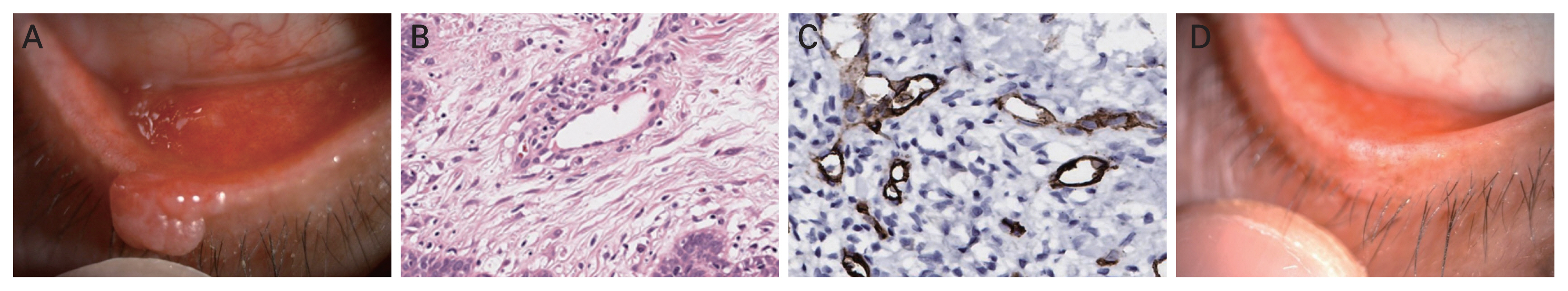
A 58-year-old man presented with a tarsal conjunctival mass involving the right lower eyelid margin, which had gradually enlarged over a period of 3 years. He had no other underlying diseases, and his best-corrected visual acuity was 20 / 20 in both eyes. A round and irregular surface mass, measuring about 0.5 cm ├Ś 0.3 cm, was observed on the tarsal conjunctiva of the right lower eyelid margin (Fig. 1A). He did not report any pain or tenderness around the mass lesion.
An excisional biopsy was conducted, and the specimen measured 0.5 cm ├Ś 0.3 cm ├Ś 0.2 cm. Histopathological examination revealed a benign spindle cell proliferative lesion with multinucleated giant cells, myxoid stroma, and superficial perivascular lymphoplasmacytic infiltration. No mitotic figures were identified. These pathological findings were consistent with conjunctival myxoid stromal tumor. On immunohistochemical staining, cells were positive for CD34 and negative for S100 and smooth muscle actin (SMA) (Fig. 1B, 1C). After the surgery, the tarsal conjunctiva and eyelid margin were clear with smooth contour (Fig. 1D).
Myxoid stromal tumors are most commonly found in the heart, skin, bones, skeletal muscles, nasal sinuses, gastrointestinal tract, and genitourinary system [1]. The occurrence of these tumors in the conjunctiva is rare. Approximately 93% of previously reported cases of conjunctival myxomas were located on the bulbar conjunctiva. There is only one case of eyelid margin-involving conjunctival myxoid stromal tumor reported by Martin et al. [2].
Immunohistochemically, conjunctival myxoid stromal tumors typically display strong positive immunoreactivity for CD34, BCL2, and vimentin, while exhibiting negative expression for S100, SOX10, and SMA. Conjunctival myxoid stromal tumors may be a presenting symptom of Carney complex, an autosomal dominant syndrome consisting of myxomas, mucocutaneous pigmentation, endocrine overactivity and melanotic schwannomas, and thus considering the potential association with serious Carney complex including atrial myxoma, it is important to include it in the differential diagnosis [3]. Hence, accurate diagnosis of conjunctival myxoid stromal tumors through immunohistochemical testing holds utmost significance.
Treatment of the conjunctival myxoid stromal tumors is simple resection. To prevent recurrence and achieve favorable cosmetic outcomes, it is important to be careful not to leave any remnants of the tumor, and to scrape the conjunctival surface smoothly. There is no report on local recurrence or malignant transformation of conjunctival myxoid stromal tumor for the past 4 months [4].
In conclusion, we present the first case of a tarsal conjunctival myxoid stromal tumor involving the eyelid margin in Korea. Although tarsal conjunctival stromal myxoid tumor is extremely rare, it should be considered as a differential diagnosis of conjunctival tumors.
 PDF Links
 PubReader
 ePub Link
 Full text via DOI
 Download Citation
 Print
Print


