Recurrence of Retinal Detachment after Scleral Buckle Removal
Article information
Abstract
Purpose
To investigate the indications for scleral buckle removal and the risk factors for the recurrence of rhegmatogenous retinal detachment after scleral buckle removal.
Methods
In this retrospective study, the medical records of all patients who underwent scleral buckle removal for the treatment of rhegmatogenous retinal detachment were reviewed.
Results
Forty eyes (40 patients) were included in this study. The indications for scleral buckle removal included exposure without infection in 23 eyes (57.5%), exposure with infection in seven eyes (17.5%), elevated intraocular pressure in six eyes (15.0%), strabismus or diplopia in three eyes (7.5%), and migration of buckle material in one eye (2.5%). After the removal of the scleral buckle, the recurrence of rhegmatogenous retinal detachment was observed in four eyes (10.0%) during follow-up, and the retina was successfully reattached after pars plana vitrectomy in all the eyes. Most clinical and ocular factors of the eyes with and without the recurrence of retinal detachment during follow-up were not different, but the eyes that underwent encircling removal were more likely to have retinal detachment recurrence during follow-up than those that underwent segmental buckle removal (n = 4 / 16 [25.0%] vs. n = 0 / 24 [0.0%]; p = 0.020).
Conclusions
Scleral buckle removal can result in the recurrence of retinal detachment. The benefits and risks of scleral buckle removal should be carefully considered before surgery, and extensive monitoring during follow-up after scleral buckle removal is important, especially for patients who underwent encircling removal.
Although pars plana vitrectomy (PPV) has been increasingly performed to treat rhegmatogenous retinal detachment (RD). However, scleral buckling (SB) remains the preferred procedure in some clinical settings, particularly for phakic eyes with a few retinal breaks located within the inferior retina. Despite the successful treatment of rhegmatogenous RD by SB, patients sometimes require the removal of buckle materials. The rates of scleral buckle removal after successful RD reattachment ranged from 1% to 24% in previous studies [1–4], and indications for removal include exposure, infection, displacement, pain, inflammation, foreign body sensation, strabismus, diplopia, and intraocular pressure (IOP) elevation [5].
The frequency of scleral buckle removal has decreased because SB for the primary repair of rhegmatogenous RD is less frequently performed than in the past [5–8]. However, scleral buckle removal can result in the recurrence of RD, which is the worst complication, although recurrent RD can be successfully repaired in most cases [9]. Known risk factors for RD recurrence include combined surgery with PPV, the presence of vitreous traction, shorter scleral buckle duration after placement, retinal tears (as opposed to holes), and unrecognized retinal breaks at the time of SB [5]. Before scleral buckle removal, the risk of RD recurrence should be balanced against the benefit of symptom relief. The purpose of this study was to investigate the indications for scleral buckle removal and the frequency of RD recurrence after scleral buckle removal. In addition, we evaluated the risk factors associated with RD recurrence after scleral buckle removal.
Materials and Methods
In this retrospective study, the medical records of all consecutive patients who underwent scleral buckle removal at Seoul National University Hospital between October 2002 and July 2019 were reviewed. Only those who showed a fully attached retina at the time of removal were included. The exclusion criteria were as follows: (1) previous SB or encircling surgery for RD due to etiologies other than rhegmatogenous, such as tractional, traumatic, and retinopathy of prematurity; (2) presence of other retinal pathologies that can cause RD or subretinal fluid formation; (3) follow-up of less than 6 months after removal.
This study was approved by the institutional review board of Seoul National University Hospital (2007-082-1141), and it was conducted following the tenets of the Declaration of Helsinki. Informed consent was waived due to the retrospective nature of the study.
The following information was obtained from a retrospective chart review: demographic characteristics; systemic disease; past ocular history; fundus findings at the time of the initial RD state, such as location and number of retinal breaks; surgical details of previous SB, such as type and materials of SB; surgical indications for scleral buckle removal; time interval between SB and the removal of the buckle; clinical courses and outcomes after scleral buckle removal. When infectious signs were observed in the case of buckle exposure, the removed materials were sent for microbial tests and culture, according to the discretion of the surgeon. In some cases, particularly for encircling, only a part of the buckle material was cut and partially removed when only that part was thought to be problematic according to the discretion of the surgeon.
The statistical analyses were performed using IBM SPSS Statistics ver. 21.0 (IBM Corp., Armonk, NY, USA). The Fisher’s exact test, Kendall’s tau-b, and the Spearman test were used. A p-value of <0.05 was considered significant. All visual acuity measurements were performed using the Snellen chart and converted to the logarithm of the minimal angle of resolution (logMAR) units for statistical analysis (finger counting = 2.0, hand motion = 2.5, light perception = 3.0, no light perception = 3.5).
Results
A total of 40 eyes in 40 patients met the study criteria and were included. The patient demographic data and clinical characteristics are summarized in Table 1. The mean age was 31.7 ± 16.3 years (range, 12 to 76 years) at the time of initial SB, and the mean time interval between placement and removal of the scleral buckle was 97.7 ± 107.0 months (range, 1 to 370 months). The mean follow-up duration after scleral buckle removal was 45.4 ± 50.0 months (range, 6 to 207 months).
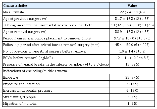
Baseline demographic and clinical characteristics of patients and eyes (n = 40)
Thirteen eyes (32.5%) underwent a 360-degree encircling using a silicone band and/or tire, and three eyes (7.5%) underwent encircling combined with segmental SB. Of 24 eyes (60.0%) that underwent segmental buckling, the scleral buckles were circumferential in 20, radial in 3, and both in 1. Two or more scleral buckles were placed in five eyes. Three eyes (7.5%) had a history of vitrectomy before SB, and 16 eyes (40.0%) underwent combined vitrectomy at the time of SB. Fourteen eyes (35.0%) required vitrectomy after SB due to failed reattachment of the retina. In total, 19 eyes (47.5%) had a previous history of vitrectomy at the time of scleral buckle removal. the mean number of vitreoretinal surgeries before scleral buckle removal was 1.6 ± 1.4 (range, 1 to 9). The mean number of retinal breaks at the time of initial SB was 1.6 ± 1.2 (range, 1 to 5), and 13 eyes (32.5%) had one or more retinal breaks within the inferior periphery between 4 and 8 o’clock. The prevalence of proliferative vitreoretinopathy or vitreous traction (n = 4 / 16 [25.0%] vs. n = 3 / 24 [12.5%]; p = 0.407) and retinal tears (n = 12 / 16 [75.0%] vs. 15 / 24 [62.5%]; p = 0.503) as causes of retinal breaks in eyes that underwent encircling and those that underwent only SB, respectively, were not different.
The mean age at the time of scleral buckle removal was 38.9 ± 18.3 years (range, 12 to 88 years). In four eyes (10.0%), the scleral buckle was removed within 6 months after placement. The indications for scleral buckle removal included exposure of the buckle without infection in 23 eyes (57.5%), exposure with infection in seven eyes (17.5%), elevated IOP in six eyes (15.0%), strabismus or diplopia in three eyes (7.5%), and migration of the buckle material in one eye (2.5%) (Table 1 and Fig. 1). The diagnosis of clinical infection was based on the observation of inflammatory signs, such as pain, redness, and purulent discharge. No intraoperative complications occurred during scleral buckle removal. Clinical infections were successfully treated with scleral buckle removal, pus drainage if required, and postoperative antibiotic eye drops. Microbial tests and cultures were performed in two eyes with clinically infected eyes, and one eye showed a positive culture for Aspergillus flavus. The patient was treated postoperatively with topical moxifloxacin and 1% voriconazole, and a granuloma persisted without infection. Among the six eyes with elevated IOP, four eyes required an Ahmed implant surgery despite scleral buckle removal, and two eyes showed normalization of IOP with medical treatment. Strabismus and diplopia were completely resolved after scleral buckle removal in two eyes, but one eye showed persistent strabismus even after strabismus surgery: 20 prism diopters of esotropia and 6 prism diopters of left hypotropia. The mean best-corrected visual acuity was 1.2 ± 1.1 logMAR (range, −0.2 to 3.5 logMAR; Snellen equivalent 20 / 320) at the time of scleral buckle removal and 1.1 ± 1.1 logMAR (range, −0.2 to 3.5 logMAR; Snellen equivalent 20 / 250) at the time of the final follow-up, and there was no significant difference (p = 0.321, Wilcoxon signed-rank test). The mean duration from the scleral buckle placement to exposure was 117.3 ± 110.7 months in eyes without infection and 157.6 ± 118.5 months in eyes with infection; the difference was not significant (p = 0.360).

Period from surgery to scleral buckle removal according to the indication of scleral buckle removal. IOP = intraocular pressure.
RD recurred after scleral buckle removal in four eyes (10.0%), and their clinical characteristics are listed in Table 2. RD recurred 15, 31, 33, and 852 days (median 32 days) after scleral buckle removal in these eyes. The median age at the time of scleral buckle removal was 41.5 years (range, 12 to 55 years), and the median duration between scleral buckle placement and removal was 20 months (range, 7 to 288 months). The indications for scleral buckle removal were strabismus or diplopia, exposure without infection, exposure with infection, and elevated IOP. Previous procedures performed were encircling in two eyes, encircling combined with segmental SB in one eye, and encircling combined with PPV in one eye. In two eyes, the encircling band was partially removed. Three parts of the encircling band were removed along with granulation tissue in one case, and a part of the encircling band was partially removed for simultaneous Ahmed implant placement and IOP control in another case. In all four eyes, only one retinal tear was observed at the time of the initial buckle surgery, and two eyes (50.0%) had a retinal break in the inferior periphery between 4 and 8 o’clock. Prophylactic laser photocoagulation was performed before scleral buckle removal in two eyes at the physician’s discretion. The recurrence of detachment was attributed to the reopening of the previous retinal break in three eyes and a new break in one eye. In an eye with a new break, RD recurrence was caused by a full-thickness macular hole 852 days after partial removal of the encircling band due to infection (Fig. 2A–2C). All patients underwent PPV for recurrent RD, and they all showed retinal reattachment at the final follow-up. On comparing the eyes with and without recurrent RD after scleral buckle removal, only the type of SB was in the groups were significantly different (Table 3). Four of 16 eyes (25.0%) that underwent encircling removal showed RD recurrence, but none of the eyes that underwent removal of only a segmental scleral buckle showed recurrence (p = 0.020).

Clinical details of four cases with retinal redetachment after scleral buckle removal

(A) Preoperative ultra-widefield fundus photography showing retinal detachment. (B,C) Horizontal and vertical B-scan optical coherence tomography images showing full thickness macular hole.
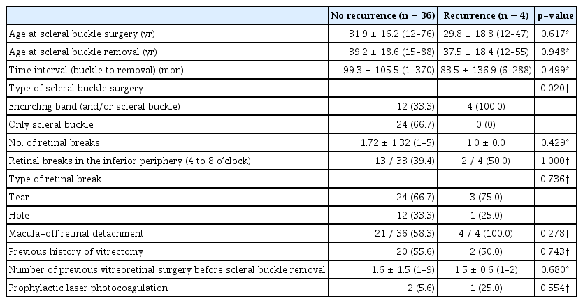
Comparison of eyes with and without recurrence of retinal detachment after scleral buckle removal
Discussion
In eyes with a stable retina, the removal of the scleral buckle that successfully reattached the retina in the primary treatment for rhegmatogenous RD raises concerns for the recurrence of RD and the necessity for additional surgery. In this study, RD recurrence was observed in 10.0% of eyes that underwent the removal of SB, which was placed as a primary treatment for rhegmatogenous RD. This was consistent with the rate of RD recurrence in recent studies, which ranged from 3.4% to 12.0% [10]. The benefits and risks of scleral buckle removal should be assessed considering the clinical situations necessitating scleral buckle removal and the known risk factors for the recurrence of RD.
The indications for scleral buckle removal were not significantly different from those in previous studies. The most common indication for scleral buckle removal was buckle material exposure, which accounted for up to 75% of cases when infectious conditions due to exposure were included. This was consistent with the findings of most previous studies on scleral buckle removal, in which exposure with or without infection constituted 47.2% to 91.8% of the cases [2,9–13]. To avoid this complication, it is important to properly cover the buckle material with the Tenon’s capsule and conjunctiva and trim the edges of the sponge or silicone tire to reduce its sharpness. In exposure cases, infection is often present, and the most common causative organism is coagulase-negative staphylococci [14]. Patients included in this study underwent scleral buckle removal almost without culture, but empirical oral antibiotics and antibiotic eye drop treatments improved the signs of infection in all the cases.
In this study, IOP elevation was the third most common cause of scleral buckle removal. The frequency of closed-angle glaucoma ranges between 1.4% and 4.4% after encircling or segmental SB [15,16], and it is caused by the forward displacement of the ciliary body due to choroidal venous congestion and the mass effect of a large explant. IOP is more likely to increase in eyes with a shallow anterior chamber, anteriorly located scleral buckle, history of encircling, high myopia, older age, and previous history of choroidal detachment [17]. In closed-angle glaucoma refractory to medical treatment, the removal of the buckle material can facilitate IOP normalization, whereas open-angle glaucoma is mostly caused by the response to corticosteroid use after surgery. In some eyes, the removal of the encircling band can also secure space for inserting a glaucoma drainage implant that would be required for the management of persistent IOP elevation even after scleral buckle removal.
Strabismus after SB is usually temporary, but persistent strabismus or diplopia occurs in approximately 5% to 25% of cases [18]. However, scleral buckle removal is reported to have a minimal effect on strabismus and diplopia caused by SB, and it does not prevent strabismus surgery [19]. In our study, one of three patients who underwent scleral buckle removal for strabismus underwent strabismus surgery, but the symptoms persisted. If persistent strabismus or diplopia exists after SB, removal may not ensure the resolution of symptoms, even after strabismus surgery.
If RD recurs after scleral buckle removal, a second surgery needs to be performed, which may affect visual acuity. In this study, retinal redetachment after scleral buckle removal involved the macula in all four eyes, but the final visual acuity was not significantly affected due to prompt and successful surgery. Redetachment occurred within 40 days, except in one case in this study, and it is important to follow up on patients frequently, particularly during the early postoperative period after scleral buckle removal. Meanwhile, long-term follow-up is also required, although the probability of late occurrence of retinal redetachment seems to be low. It is of interest that a case with late recurrence of RD was associated with a new retinal break rather than reopening of preexisting retinal breaks. In particular, more attention should be paid to cases that underwent encircling removal, as shown by the higher risk of retinal redetachment in this study.
Compared with the removal of the segmental buckle, the removal of the encircling band was associated with a higher risk of RD in this study. After segmental SB, the height of the buckle decreased over a long period. Serial A-scan ultrasonography measurements of segmental buckle heights in a prospective study showed that only half of the buckles maintained their original heights [20]. Although there are no reports on the long-term change in scleral buckle height after encircling, the encircling height is likely not to decrease over time because of the presence of the Watzke sleeve for securing both ends of the silicone band and adjusting the tension in the band. It can be hypothesized that the eyes that remained attached after segmental buckle surgery, even after a decrease in buckle height during follow-up, may have had decreased vitreous traction on the retinal breaks compared to the time of SB, which can be supported by the decreased buckle height. In these eyes, the removal of the segmental buckle may not cause the reopening of the retinal breaks. On the other hand, the removal of the encircling band may have more influence on the reopening of the retinal breaks if the eyes have unchanged vitreous traction on the retinal breaks, which needs to be supported by the sufficient height of encircling.
This study has some limitations. First, it was a retrospective study with a small sample. Second, the follow-up lasted for as short as 6 months in some eyes, and RD recurrence during the later stage may have been missed, biasing the rate of retinal redetachment. Although most redetachments were reported to occur within 3–6 months after scleral buckle removal [10,11], redetachment can occur even 2 years after scleral buckle removal, as observed in the present study. Third, the number of SB cases for rhegmatogenous RD during the study period was not included in the analysis, and the frequency of scleral buckle removal could not be obtained in this study. In a recent study on scleral buckle removal performed between 2004 and 2013, the overall rate was 5.7% [10]. Despite these limitations, this study has shown that the indications for scleral buckle removal and the probability of retinal redetachment were comparable to those of previous studies performed within the era of vitrectomy. The benefits and risks of scleral buckle removal should be carefully considered before surgery, and caution should be exercised during the early postoperative period after scleral buckle removal, especially for patients who undergo encircling removal.
Notes
Conflict of Interest
No potential conflict of interest relevant to this article was reported.