 |
 |
| Korean J Ophthalmol > Volume 34(2); 2020 > Article |
|
Abstract
Purpose
We report the clinical outcomes of retinal capillary hemangioma (RCH) after the application of various treatments.
Methods
We performed a retrospective chart analysis of eight eyes treated for RCH between August 2009 and January 2018. During the follow-up period, the status and progression of the RCHs were checked by fundus photography, fluorescein angiography, and optical coherence tomography, and additional treatments were applied when necessary.
Results
Three of the five patients had bilateral RCH, and two had unilateral RCH. Six eyes received laser photocoagulation; two eyes received cryotherapy, and one eye received intravitreal Avastin injection. Three eyes each had intravitreal triamcinolone injection, subtenon triamcinolone injection, and intravitreal dexamethasone injection to control inflammation. Also, two patients took oral prednisolone, and one patient used prednisolone eye drops to control inflammation. Two eyes underwent vitrectomy and scleral buckling due to deterioration of the epiretinal membrane and vitreal traction, respectively. As a result of those treatments, the tumors were stable in five of the eight eyes. However, one eye is now in a pre-phthisis state, and one patient who refused treatment showed progression of the tumor, epiretinal membrane, and traction.
Conclusions
Because RCHs vary in size, the degree of inflammation, and symptoms, this disorder should be actively treated on a case-by-case basis. Fluorescein angiography should be used periodically to determine recurrence of the tumor or inflammation, and the appropriate treatment should be repeated as necessary. Moreover, regular systemic screening tests for von Hippel-Lindau disease should be performed in RCH patients to ensure that they have no abnormalities other than in the eye.
Retinal capillary hemangioma (RCH) is a vascular hamartoma that can be unrelated to systemic symptoms or occur as part of von Hippel-Lindau disease (VHL), an autosomal dominant hereditary disease caused by a partial defect in the short arm of chromosome 3 that can cause subtentorial hemangioblastoma, renal cancer, pheochromocytoma, and RCH [1,2].
RCH is a red/pinkish round mass that occurs in the periphery of the retina or around the optic disc; its exudative appearance is similar to Coats' disease, but the tumor is fed by a dilated, tortuous retinal artery and drained by an engorged vein. The tumor can be also accompanied by vitreoretinal traction, vitreous hemorrhage, or tractional retinal detachment [3,4,5,6,7]. The decrease of visual acuity seen in RCH is mainly caused by subretinal or intraretinal fluid due to exudation from the proliferating tumor, an epiretinal membrane (ERM), or tractional retinal detachment [7,8,9,10].
The treatment of RCH is based on tumor size, location, and secondary complications [11,12]. The patient can be observed without treatment or treated with laser therapy, cryotherapy, photodynamic therapy with verteporfin [7,13,14,15], or vitrectomy [10,16,17,18,19,20].
In the present study, we report the clinical outcomes of RCH after appropriate treatment, including observation, laser photocoagulation, cryotherapy, intravitreal triamcinolone injection, or surgery.
We conducted a retrospective chart review of eight eyes with RCH treated between August 2009 and January 2018. Diagnosis was based on the typical fundus findings of RCH and fluorescein angiography. All patients underwent the comprehensive ocular examination, including best-corrected visual acuity (BCVA), intraocular pressure (IOP), slit-lamp examination, and fundus examination. Fundus photography, fluorescein angiography, and optical coherence tomography (OCT) were routinely performed to assess disease progression in the affected eye and check for disease onset in the opposite eye. Family and individual medical histories were also examined. This study adhered to the tenets of the Declaration of Helsinki and was approved by the institutional review board of Chungnam National University Hospital in the Republic of Korea (2018-08-026). Informed consent was waived due to the retrospective nature of the study.
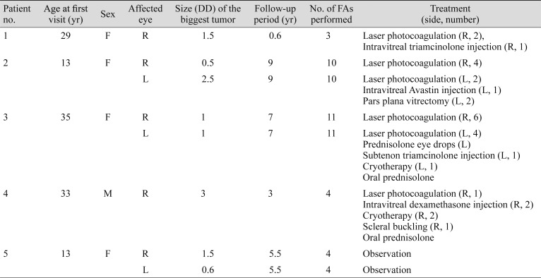
The basic informations of the patients are presented in Table 1. We evaluated a total of eight eyes of five patients who were diagnosed with RCH. The mean patient age was 24.6 ± 10.8 years. There were five affected right eyes and three affected left eyes.
A 29-year-old female patient visited our clinic with a 3-month history of a floater in the right eye. At the first visit, the BCVA in both eyes was 20 / 20, and the IOP was normal. There was no relevant family history, ocular trauma history, or other systemic problems. There were also no abnormal findings in the anterior segment examination. But on the fundus examination, +2 inflammatory cells were observed in the anterior vitreous cavity of the right eye, and three RCHs in the superotemporal area of the retina, vitreous traction, and exudates were also observed (Fig. 1A, 1B). The size of the largest RCH in the right eye was 1.5 disc diameters. We performed laser photocoagulation for the tumor (0.3 seconds, 275 mW) and administered 4 mg/0.1 mL intravitreal triamcinolone acetonide (Dong Kwang, Seoul, Korea). To diagnose VHL, we requested medical examinations by a neurosurgeon and urologist, but no abnormalities were found. Two weeks later, additional laser photocoagulation was performed on the right eye. After 2 weeks, 1 month, and 4 months, the patient visited our clinic for follow-up observations, and the tumor remained stable until last follow-up (Fig. 1C, 1D).
A 13-year-old female patient visited our hospital due to decreased visual acuity in the left eye. The BCVA of the left eye was 20 / 100, and the BCVA of the right eye was 20 / 15. The IOP, findings of the eyelid, cornea, and conjunctiva were all normal, and 1+ inflammatory cells were observed in the anterior chamber of both eyes. In the fundus examination, an RCH with 2.5 disc diameters was observed in the left eye with dilated retinal vessels and exudates, and macular edema was detected by OCT (Fig. 2A-2C). Fluorescein angiography of the right eye showed a localized leakage of fluorescence. Bevacizumab (Avastin; Genentech, San Francisco, CA, USA) was injected intravitreally to treat the macular edema in the left eye, and laser photocoagulation was performed (0.3-0.5 seconds, 150-180 mW) in the left eye. Kidney and brain imaging tests were performed for VHL diagnosis, and no abnormalities were found. After 2 months, the macular edema of the left eye improved, and laser photocoagulation was applied to both eyes.
One month later, ERM with vitreous traction was observed in the left eye (Fig. 2D, 2E). Then we recommended a pars plana vitrectomy. The patient received a pars plana vitrectomy and silicone oil injection at another hospital. One year later, fluorescein angiography of the right eye showed an increase in fluorescein leakage, and additional laser photocoagulation was performed. Four years after surgery, silicone oil was observed in the anterior chamber of the left eye, and the oil was removed at another hospital because of high IOP. After 6 weeks, silicone oil was injected again. At the last visit, the left eye was in a pre-phthisis state, and the right eye was stable after two additional laser photocoagulation treatments (Fig. 2F).
A 35-year old female patient visited our hospital with vision loss in the left eye, which had started 1 week before. She had previously been diagnosed with glaucoma in the left eye and was being treated with 1% brinzolamide/0.5% timolol maleate (Elazop; Alcon, Fort Worth, TX, USA). The BCVA was 20 / 50 in the left eye and 20 / 15 in the right eye. The IOP was 29 mmHg in the left eye and 18 mmHg in the right eye. There were 1+ inflammatory cells in the anterior chamber of both eyes. On the fundus examination, anterior vitreous cells were trace in the right eye and 2+ in the left eye. RCHs with engorged vessels were observed at the temporal area of the right eye and the inferotemporal periphery of the left eye (Fig. 3A, 3B). Laser photocoagulation was performed (0.2 seconds) in the left and right eyes (300 and 190 mW, respectively). To control the inflammation, 1% prednisolone acetate (Pred Forte; Allergan, Irvine, CA, USA) was administered to the left eye, in addition to 30 mg of oral prednisolone for 1 week. The patient was referred to the hematology and oncology departments for a VHL evaluation, and no additional abnormality was found. After 1 month, the exudates increased, and fluorescein leakage was still observed around the RCH in the left eye during fluorescein angiography. The tumor was still in the active state, and laser treatment was considered insufficient because the tumor was located in the far periphery of the temporal retina. Triamcinolone (4 mg/0.1 mL) was injected into the subtenon, and cryotherapy was performed in the left eye. The patient underwent additional laser photocoagulation in both eyes when the fluorescein leakage increased during fluorescein angiography. After a total of two laser photocoagulations in the left eye and four in the right eye, the patient enjoyed a stable state for 6 months to the last follow-up (Fig. 3C, 3D).
A 33-year-old male visited our clinic with a floater in the left eye and decreased visual acuity; he was diagnosed with VHL. In 2006, he underwent craniotomy and tumor resection for right ventricular angioblastoma. He had a relevant family history, with his father receiving surgery for a tumor of the spine. At the time of the first visit, the BCVA was 20 / 20 in both eyes. No specific finding was noted in the IOP and anterior segment examinations.
During the fundus examination, an RCH was found, along with a dilated vein on the temporal retina of the right eye. Fluorescein angiography showed hyperfluorescence of the whole retina due to inflammation (Fig. 4A-4D). We injected intraocular dexamethasone (Ozurdex, Allergan) to control inflammation and performed laser photocoagulation (0.25 seconds, 250 mW) one week later. Two weeks after that, increased vascular dilatation and retinal hemorrhage around the tumor were observed, along with vitreous opacity. Cryotherapy was performed because additional laser photocoagulation was deemed insufficient to control tumor progression. Oral prednisolone (20 mg for 1 week followed by 10 mg for 1 week) was administered, and ophthalmic examinations were conducted every month. Three months later, fluorescein leakage in the RCH was observed during fluorescein angiography of the right eye, so we assumed that the tumor was still in the active phase. A second injection of intraocular dexamethasone was performed, and a second cryotherapy was performed 2 weeks later in the right eye. However, the temporal retinal traction deteriorated, so scleral buckling was performed on the right eye (Fig. 4E-4H). After surgery, the tumor was stable for 2.5 years without further treatment (Fig. 4I-4L).
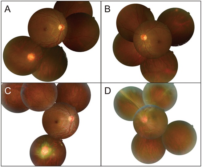
A 13-year-old female patient visited our hospital after a pediatric consultation for VHL. Her mother had a history of cerebral hemorrhage of the right middle cerebral artery, and her sister underwent a follow-up observation at our eye and pediatric clinics for VHL. There were no ophthalmic symptoms at the first visit. The BCVA was 20 / 20 in both eyes; the IOP was normal, and there were no abnormal findings in the anterior segment examinations. On the fundus examination, RCHs and dilated blood vessels were observed in the inferotemporal side of the retina in the right eye and the superotemporal retina in the left eye (Fig. 5A, 5B). She was also diagnosed with VHL following a genetic test administered at the pediatric clinic. We recommended laser photocoagulation, but her parents denied treatment, preferring to observe without any treatment. Nine years after the first visit, the patient complained of decreased visual acuity in the left eye. The BCVA was 10 / 20, and retinal traction with ERM was observed. We explained that pars plana vitrectomy and cryotherapy would be needed, but the patient rejected the treatment for financial reasons. After 2 months, the BCVA was 20 / 60, and retinal traction with ERM had progressed (Fig. 5C, 5D). We again explained the need for surgical treatment, but the patient refused treatment and was referred to another hospital closer to her hometown to enable easier observation.
RCH is an uncommon hemangioma of the retina that can occur independently or in association with VHL [1]. RCH is the most common tumor and early finding of VHL [21]. Singh et al. [22] reported that with capillary hemangioma alone, the probability of being later diagnosed with VHL was 25%. Therefore, patients with RCH should be screened regularly for systemic abnormalities outside the eye [23]. Most RCHs progressively increase in size, and an increase in subretinal fluid can produce total retinal detachment and hemorrhage in the vitreous, retina, or subretinal space, resulting in decreased visual acuity [7]. Therefore, early diagnosis and proper treatment of RCHs are important to maintain visual acuity [6]. In general, the treatment of RCH depends on the size of the tumor, its location, and associated findings such as the presence of subretinal fluid and retinal traction [1]. If the tumor is small or in the far periphery, the patient can be observed without treatment. Otherwise, various forms of treatment are available, including laser therapy, cryotherapy, photodynamic therapy, and vitrectomy. However, all those treatments can cause complications such as a temporary increase in the exudate or the formation of an ERM [10,24]. Therefore, treatment should be selected properly according to the patient's individual situation. We retrospectively reviewed the clinical course of eight eyes diagnosed with RCH, and we found that when an appropriate treatment modality was used, the RCH was stabilized.
Laser photocoagulation is the most widely used treatment modality when the tumor is small and not located in the far periphery [7,25]. The purpose of laser photocoagulation is to reduce the diameter of the feeding and draining vessels and exudates. With a one disc diameter, complete degeneration is histopathologically possible, and clinical improvement can be seen in tumors up to 2.5 disc diameters (<4.5 mm) [26,27]. The laser should irradiate the tumor directly with a large-diameter beam and long irradiation time (0.2-0.5 seconds) so that the entire tumor turns white. We administered intravitreal triamcinolone injections to control inflammation along with laser photocoagulation at the time of diagnosis for a 1.5-disc diameter RCH in the right eye in case 1. We then performed additional laser photocoagulation as necessary. Inflammation of the lesion and the peripheral retina was stable for approximately 4 months. In case 2, RCHs of less than 0.5 disc diameter were seen in the right eye, so laser photocoagulation was performed. Additional laser photocoagulation was performed to stabilize the lesions every time fluorescence leakage increased in the regular fluorescein angiography, and the lesions remained stable until the last follow-up. Laser photocoagulation has the advantage of being repeatable and additive, that is, it can be given at a weak power several times, to reduce the possibility of inflammation. If abnormal findings, such as fluorescein leakage on fluorescein angiography, are observed during periodic follow-up, additional laser treatment should be performed, followed by observation.
However, if the tumor is relatively large, is located in the far periphery, or is accompanied by exudative retinal detachment, laser treatment can be ineffective, and cryotherapy should be considered [28,29]. The double freeze-thaw technique is recommended to prevent proliferative vitreoretinopathy, and it should be applied until the ice ball completely encloses the RCH [30,31]. Case 3 in this study had RCHs in both eyes, and inflammation was observed in the anterior chamber of both eyes. Laser photocoagulation, oral prednisolone, and eye drops were used to control the inflammation. However, the laser treatment was insufficient in the left eye because the inflammation persisted and the tumor was not stable. After a subtenon steroid injection and cryotherapy, a stable condition was achieved. In cases with a relatively large RCH, laser treatment or cryotherapy should be applied aggressively to slow the progression of the lesion and prevent deterioration. However, as with laser photocoagulation, cryotherapy can cause a transient increase in exudate or proliferative vitreoretinopathy [32]. Also, cryotherapy is an invasive and destructive treatment modality that requires an incision in the conjunctiva, and it is usually accompanied by steroid therapy to control inflammation. In case 4, because the lesion was located in the far periphery of the right eye, cryotherapy was used instead of laser photocoagulation, but peripheral retinal traction occurred later. Although pars plana vitrectomy could be conducted in such cases, that patient was treated with scleral buckling because of his young age and possible postoperative complications after vitrectomy. That tumor remained in a stable state at the last follow-up.
Recent studies indicate that RCH might depend on vascular endothelial growth factor (VEGF) because patients with VHL who present with RCH have elevated ocular levels of anti-VEGF [33]. Anti-VEGF therapy has been reported to reduce vascular permeability, and many RCH cases have been treated successfully with intravitreal bevacizumab injections. Repeated intravitreal injections of bevacizumab stopped the growth of an existing RCH and perhaps inhibited the development of new hemangiomas in the treated eye [34,35]. Bevacizumab treatment is less likely to promote cataract development than steroid injections, making it useful for young adults. However, the duration of its effect is short, and if the inflammation or macular edema is severe, subtenon or intravitreal steroid injection is more effective.
Photodynamic therapy with verteporfin can also be considered; it is known to effectively penetrate the subretinal fluid and can be used instead of laser photocoagulation to treat large tumors [36,37,38]. Moreover, complications such as vitreous hemorrhage and proliferative vitreoretinopathy are less common with photodynamic therapy than with cryotherapy [36]. Combining photodynamic therapy with intravitreal bevacizumab injections is also an effective treatment for RCH. However, there have been reports of severely decreased visual acuity caused by extensive exudative retinal detachment after that combination therapy [24]. Furthermore, because photodynamic therapy is too expensive to be repeated, it can be done too strongly. Therefore, it should be selected only when the specific situation warrants it.
In general, pars plana vitrectomy is not the primary treatment for small, peripheral RCHs, but it is considered after laser treatment or cryotherapy in cases with significantly decreased visual acuity caused by a refractory tumor or in the presence of ERM, macular edema, and tractional and serous retinal detachment [16,17,18]. Case 2 patient had RCHs in both eyes and was treated with laser photocoagulation, but the tumor in the left eye, of 2.5 disc diameters, progressed. Thus, we performed pars plana vitrectomy and silicone oil injection to treat the proliferation of exudates and ERM. Because silicone oil was observed in the anterior chamber and the IOP was high in the left eye, silicone oil reinjection was performed after silicone oil removal. Although the patient underwent pars plana vitrectomy due to the lack of tumor regression after laser treatment, her condition did not improve. Like the other treatments, pars plana vitrectomy risks some complications, such as hemorrhage because of difficulty in controlling the bleeding from the feeder and drainage vessels around the tumor or phthisis, as in case 2. Therefore, surgery is usually performed to simply remove the ERM or treat the retinal detachment. Controversy remains regarding the best time for surgical treatment of ERMs caused by RCH. Natural regression due to complete posterior vitreous detachment can occur after photocoagulation therapy. However, a recurrence of ERM can be also seen, even after surgical treatment [17]. In case 5, when RCH was diagnosed, the patient underwent regular observation without any treatment. After 9 years of observation, ERM occurred and progressed rapidly with traction. If a lesion progresses rapidly in this manner, removal of the ERM and traction through vitrectomy could result in a better visual prognosis. RCHs vary in size and the degree of inflammation. Ophthalmologists should consider not only patient symptoms but also findings from the fundus examination, OCT, and fluorescein angiography. Fluorescein angiography should be performed routinely during the follow-up period to determine the presence of fluorescence leakage around the tumor. It is a useful method for checking the occurrence of new lesions or the recurrence of an angioma after proper treatment. In particular, clinicians should confirm that inflammation in the eye is well-controlled by checking for fluorescence staining in the peripheral area. In the case of inflammation caused by a tumor or treatment, steroid injections might be necessary. In cases 1, 3, and 4 in this study, steroid injections before and after laser photocoagulation or cryotherapy regulated the inflammation and yielded stable results.
A limitation of this study is the small number of patients included. However, RCH is a rare disease and has been insufficiently reported in the Republic of Korea. Therefore, we have reported RCH cases in which relatively stable results were achieved using various appropriate treatments.
In conclusion, because RCHs vary in size and degree of inflammation at the time of diagnosis, the treatment modalities should be adjusted to meet the needs and condition of each patient, and it is important to administer appropriate treatment repeatedly over a long period of time to control tumor progression and prevent tumor recurrence. Also, it is necessary to control the inflammation caused by the tumor or treatment, and steroid injections should be considered. During the follow-up period, patients should be checked for recurrence by means of regular fluorescein angiography, and laser photocoagulation or cryotherapy should be repeated as necessary. Additionally, because RCH is the most common and early finding of VHL, patients with RCH require periodic screening for VHL using regular systemic tests.
Notes
Conflict of Interest: No potential conflict of interest relevant to this article was reported.
REFERENCES
2. Lamiell JM, Salazar FG, Hsia YE. von Hippel-Lindau disease affecting 43 members of a single kindred. Medicine (Baltimore) 1989;68:1-29.


3. Shields JA, Shields CL. Intraocular tumors: an atlas and textbook. 2nd ed. Philadelphia: Lippincott Williams & Wilkins; 2008. p. 368-369.
4. Maher ER, Yates JR, Harries R, et al. Clinical features and natural history of von Hippel-Lindau disease. Q J Med 1990;77:1151-1163.


5. Webster AR, Maher ER, Moore AT. Clinical characteristics of ocular angiomatosis in von Hippel-Lindau disease and correlation with germline mutation. Arch Ophthalmol 1999;117:371-378.


6. Ridley M, Green J, Johnson G. Retinal angiomatosis: the ocular manifestations of von Hippel-Lindau disease. Can J Ophthalmol 1986;21:276-283.


7. Annesley WH Jr, Leonard BC, Shields JA, Tasman WS. Fifteen year review of treated cases of retinal angiomatosis. Trans Sect Ophthalmol Am Acad Ophthalmol Otolaryngol 1977;83:OP446-OP453.

8. Singh AD, Damato BE. Clinical ophthalmic oncology: basic principles. Cham: Springer; 2019. p. 99-105.
9. Lee DS, Kim YY, Kim SY, Kim SD. Treatment of retinal detachment occurring in two cases of von Hippel-Lindau syndrome. J Korean Ophthalmol Soc 2001;42:1636-1640.
10. Baek SK, Jin SY, Lee YH, Lee TG. A case report of primary vitrectomy in multiple bilateral retinal capillary hemangiomas accompanying epiretinal membrane. J Korean Ophthalmol Soc 2014;55:928-935.

11. Whitson JT, Welch RB, Green WR. Von Hippel-Lindau disease: case report of a patient with spontaneous regression of a retinal angioma. Retina 1986;6:253-259.


13. Sachdeva R, Dadgostar H, Kaiser PK, et al. Verteporfin photodynamic therapy of six eyes with retinal capillary haemangioma. Acta Ophthalmol 2010;88:e334-e340.


14. Fong AH, Li KK, Wong D. Intravitreal ranibizumab, photodynamic therapy, and vitreous surgery for the treatment of juxtapapillary retinal capillary hemangioma. Graefes Arch Clin Exp Ophthalmol 2011;249:625-627.


15. Kremer I, Gilad E, Ben-Sira I. Juxtapapillary exophytic retinal capillary hemangioma treated by yellow krypton (568 nm) laser photocoagulation. Ophthalmic Surg 1988;19:743-747.


16. Gaudric A, Krivosic V, Duguid G, et al. Vitreoretinal surgery for severe retinal capillary hemangiomas in von hippel-lindau disease. Ophthalmology 2011;118:142-149.


17. McDonald HR, Schatz H, Johnson RN, et al. Vitrectomy in eyes with peripheral retinal angioma associated with traction macular detachment. Ophthalmology 1996;103:329-335.


18. Johnson MW, Flynn HW Jr, Gass JD. Pars plana vitrectomy and direct diathermy for complications of multiple retinal angiomas. Ophthalmic Surg 1992;23:47-50.


19. Chew EY, Schachat AP. Capillary hemangioblastoma of the retina and von Hippel-Lindau disease. In: Ryan SJ, Retina. 5th ed. London: Saunders/Elsevier; 2013. p. 2156-2163.
20. Lee HJ, Kang SH, Kim HC. Three cases of retinal capillary hemangioma presenting with retinal detachment. J Korean Ophthalmol Soc 2003;44:1936-1942.
22. Singh A, Shields J, Shields C. Solitary retinal capillary hemangioma: hereditary (von Hippel-Lindau disease) or nonhereditary? Arch Ophthalmol 2001;119:232-234.


23. Hinz BJ, Schachat AP. Capillary hemangioma of the retina and von Hippel-Lindau disease. In: Ryan SJ, Hinton DR, Schachat AP, , Retina. 4th ed. St. Louis: Mosby; 2006. p. 615-624.
24. Kim HM, Park KH, Woo SJ. Massive exudative retinal detachment following photodynamic therapy and intravitreal bevacizumab injection in retinal capillary hemangioma. Korean J Ophthalmol 2015;29:143-145.


25. Schmidt D, Natt E, Neumann HP. Long-term results of laser treatment for retinal angiomatosis in von Hippel-Lindau disease. Eur J Med Res 2000;5:47-58.

26. Shields JA. The expanding role of laser photocoagulation for intraocular tumors. The 1993 H. Christian Zweng Memorial Lecture. Retina 1994;14:310-322.


27. Lane CM, Turner G, Gregor ZJ, Bird AC. Laser treatment of retinal angiomatosis. Eye (Lond) 1989;3:33-38.


29. Shields JA. Response of retinal capillary hemangioma to cryotherapy. Arch Ophthalmol 1993;111:551


30. Welch RB. Von Hippel-Lindau disease: the recognition and treatment of early angiomatosis retinae and the use of cryosurgery as an adjunct to therapy. Trans Am Ophthalmol Soc 1970;68:367-424.


31. Singh AD, Nouri M, Shields CL, et al. Treatment of retinal capillary hemangioma. Ophthalmology 2002;109:1799-1806.


32. Watzke RC. Cryotherapy for retinal angiomatosis: a clinicopathologic report. Arch Ophthalmol 1974;92:399-401.


33. Los M, Aarsman CJ, Terpstra L, et al. Elevated ocular levels of vascular endothelial growth factor in patients with von Hippel-Lindau disease. Ann Oncol 1997;8:1015-1022.


34. Ach T, Thiemeyer D, Hoeh AE, et al. Intravitreal bevacizumab for retinal capillary haemangioma: longterm results. Acta Ophthalmol 2010;88:e137-e138.


35. Chelala E, Dirani A, Fadlallah A. Intravitreal anti-VEGF injection for the treatment of progressive juxtapapillary retinal capillary hemangioma: a case report and mini review of the literature. Clin Ophthalmol 2013;7:2143-2146.


36. Atebara NH. Retinal capillary hemangioma treated with verteporfin photodynamic therapy. Am J Ophthalmol 2002;134:788-790.


Fig. 1
Clinical outcome of a case 1 patient. (A) Fundus photographs and (B) fluorescein angiography of the right eye of a 29-year-old patient at the initial visit. Three round, orange retinal masses <1.5 disc diameters with dilated feeder vessels and exudations were observed in the peripheral retina. Leakage of fluorescence associated with the lesions was revealed by fluorescein angiography. (C) The size of the masses and exudates decreased after two laser photocoagulation treatments. (D) Leakage around the lesions also decreased after two laser photocoagulation treatments.
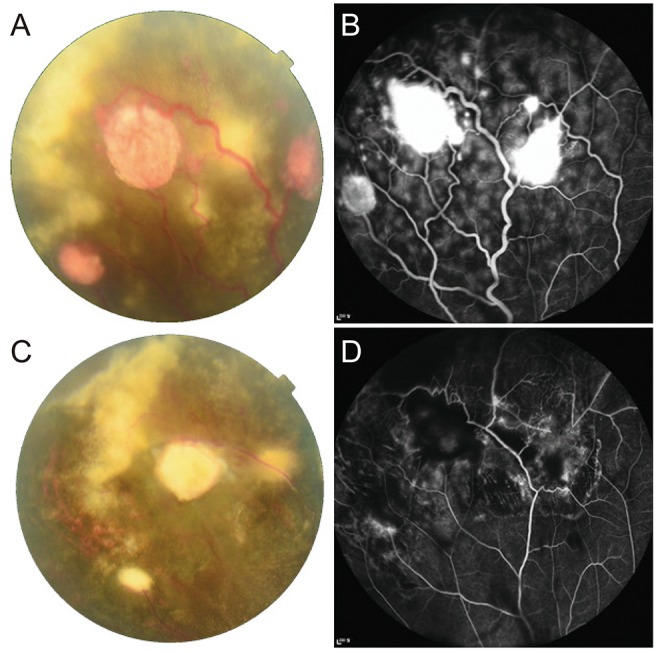
Fig. 2
Clinical outcome of a case 2 patient. (A,B) Fundus photographs and (C) fluorescein angiography (FA) of the left eye of a 13-year-old patient before treatment. A tumor of 2.5 disc diameters with dilated vessels was found in the temporal retina, and exudates were deposited around the vessels. Leakage of fluorescence associated with the lesions was revealed by FA. Although laser photocoagulation was repeated, (D) fundus photographs and (E) optical coherence tomography showed vitreous traction with an epiretinal membrane, and tumor progression was not completely attenuated. (F) The retinal masses in the right eye were stable on FA after four laser photocoagulation treatments.

Fig. 3
Clinical outcome of a case 3 patient. (A) Fundus photograph and (B) fluorescein angiography (FA) of the left eye of a 35-year-old patient before cryotherapy. There were exudations with dilated feeder vessels, and (B) FA revealed leakage from the lesion. Seven years after cryotherapy and additional laser photocoagulation treatments, a (C) fundus photograph and (D) FA showed no dilated vessels, exudates, or leakage around the mass.
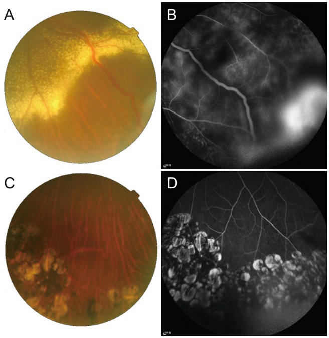
Fig. 4
Clinical outcome of a case 4 patient. (A) The fluorescein angiography (FA) of the right eye of a 33-year-old patient at the first visit revealed inflammation throughout the retina. (B) Fundus photography, (C) FA, and (D) optical coherence tomography showed a mass of 3 disc diameters before treatment. After two cryotherapy treatments, (E) the inflammation disappeared in the FA, but retinal traction worsened in (F) the fundus photograph, (G) FA, and (H) optical coherence tomography. (I-L) After scleral bucking, the right eye reached a stable state.
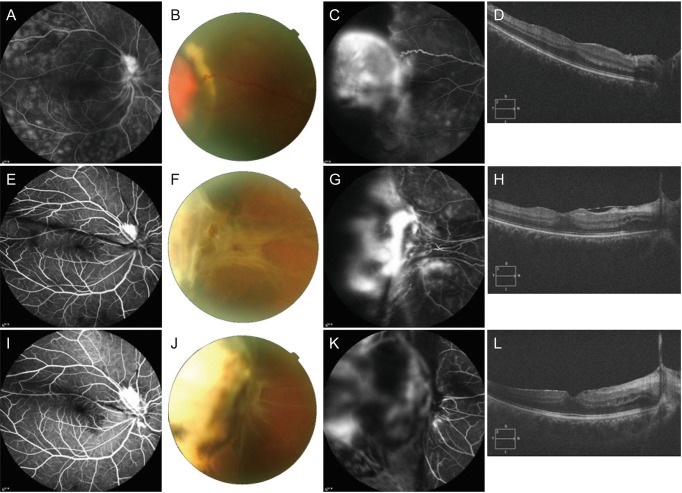
Fig. 5
Clinical outcome of a case 5 patient. (A,B) Fundus photographs of a 13-year-old patient at the initial visit. The retinal tumors were in the right inferotemporal peripheral retina and the left superotemporal retina. Nine years later, without any treatment, (C) the tumor in the right eye became enlarged, and (D) retinal traction and ERM had progressed in the left eye.
- TOOLS
-
METRICS

- Related articles
-
Clinical features of strabismus in patients with congenital optic disc anomaly2021 April;35(2)
Clinical Results of Anti-adhesion Adjuvants after Endonasal Dacryocystorhinostomy2018 December;32(6)
Outcomes of Cataract Surgery Following Treatment for Retinoblastoma2017 February;31(1)


 PDF Links
 PubReader
 Full text via DOI
 Full text via PMC
 Download Citation
 Print
Print


