

Dear Editor,
Ocular features that can accompany vitiligo include hypopigmented spots on the iris, retinal pigment epithelium (RPE) hypopigmentation, uveitis, and diffuse choroidal hypopigmentation [1]. According to a dermatopathology textbook, the ŌĆ£essential process in vitiligo is the destruction of melanocytesŌĆØ [2]. Choroidal vitiligo might occur as a primary or secondary process; primary choroidal vitiligo can occur as an idiopathic condition without preceding inflammation, and secondary choroidal vitiligo might occur after intraocular inflammation, most often in Vogt-Koyanagi-Harada syndrome. The pathogenesis of Vogt-Koyanagi-Harada syndrome is related to aberrant T cell-mediated immune response directed against self-antigens expressed by melanocytes. Clinical presentation correlates with the destruction of melanocytes in affected areas such as the eyes, ears, skin, and central nervous system [3]. Here, we report a case of atypical primary ocular vitiligo with X-shaped choroidal hypopigmentation.
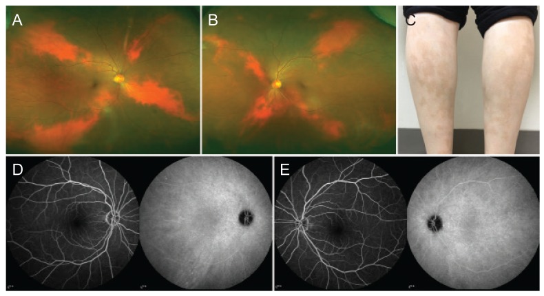
A 55-year-old Asian woman was referred to the ophthalmology clinic because of abnormal fundus appearance. Her best-corrected visual acuity was 20 / 32 in the right eye and 20 / 20 in the left eye. Slit-lamp examination was completely normal with the exception of the fundus. Both fundi showed X-shaped areas of choroidal hypopigmentation (Fig. 1A, 1B). The patient reported having cutaneous vitiligo of the lower extremities (Fig. 1C). Tiny scattered hypofluorescent spots were present on fluorescein angiography and hyperfluorescent spots were observed on indocyanine green angiography bilaterally (Fig. 1D, 1E). The ocular features involved only choroidal vitiligo without RPE abnormalities. She had no history of general or ocular disease except for cutaneous vitiligo. Six months after the diagnosis of ocular vitiligo, there was no intraocular inflammation or progression of choroidal hypopigmentation.
In the ophthalmology literature, ocular vitiligo usually presents with diffuse choroidal vitiligo with uveal inflammation [4]; X-shaped choroidal hypopigmentation is a relatively rare presentation. In this case, the coexistence of cutaneous and ocular vitiligo may be explained by the embryological origin of the affected cells. Both skin and choroidal melanocytes are commonly derived from neural crest cells. Alternately, choroidal hypopigmentation might have been congenital and the coexistence of cutaneous vitiligo in our patient might have been unrelated.
In conclusion, we described an atypical pattern of ocular vitiligo presenting with cutaneous vitiligo in Asian women. This asymptomatic choroidal hypopigmentation allows for normal visual acuity and normal retinal and RPE anatomy.
 PDF Links
 PubReader
 Full text via DOI
 Full text via PMC
 Download Citation
 Print
Print


