

Dear Editor,
A mucocele is composed of a respiratory epithelial lined mucus-containing lesion with accumulation of mucoid secretion. Sometimes, it produces remodeling of the bony structure and influences the adjacent tissues [1]. In this case, extension of the ethmoid sinus mucocele and remodeling of the orbital structure were major causes of acquired Brown syndrome. Although various causes of acquired Brown syndrome have been described, this is the first reported case of acquired Brown syndrome due to ethmoidal sinus mucocele in Korea.
A 69-year-old male presented to our institution with a newly-developed diplopia. There was no history of trauma or any medical illness. At initial examination, visual acuity was 20 / 30 in both eyes. The intraocular pressure was 17 mmHg in the right and 20 mmHg in the left eye. There was no relative afferent pupillary defect and color vision was normal.
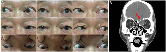
A soft compressible tender mass measuring 2 × 2 mm on the superonasal aspect of the left upper lid extending from the trochlea to the medial canthal tendon was noted. A motility examination showed an underelevation in adduction of the left eye mimicking Brown syndrome (Fig. 1A). A left hypotropia of 2 prism diopters (PD) to 4 PD in primary gaze, which increased to 6 PD in right and upgaze, was present. Forced duction testing indicated a restriction of elevation in adduction of the left eye. Computed tomography (Fig. 1B) revealed an expansile nonenhancing 30 × 25 mm mass of the left ethmoidal sinus having an intraorbital extension consistent with a mucocele. The mass appeared to be pressing on the globe causing effacement of the left medial rectus and superior oblique tendon with anterolateral displacement of the trochlea. The patient underwent endoscopic ethmoidectomy and evacuation of the mucocele by an otolaryngologist. Postoperatively at two months, there was complete resolution of the diplopia and the patient was asymptomatic. A forced duction test revealed no restrictions.
Brown syndrome is characterized by limited elevation in adduction with positive forced duction testing [2]. The mechanism of acquired Brown syndrome in this case was thought to be the ethmoidal sinus mass preventing anterior tendon movement through the trochlea or the spread of inflammation from an adjacent sinus disease [3]. Complete resolution of symptoms after surgery suggests that mechanical effects were the main cause in this case. Although acquired Brown syndrome is rare, ethmoidal sinus mucocele should be considered in differential diagnosis of acquired Brown syndrome.
 PDF Links
 PubReader
 Full text via DOI
 Full text via PMC
 Download Citation
 Print
Print


