Conjunctival Hypertrophic Scar Following Cryotherapy for Retinopathy of Prematurity
Article information
Abstract
A 6-year-old boy was referred to our hospital with symblepharon and lateral canthal deformity in both eyes, which developed 6 years ago. The patient was born at 27 weeks gestation. He had received cryotherapy for retinopathy of prematurity. One month after cryotherapy, he developed a conjunctival scar with symblepharon in both eyes and underwent symblepharon lysis at another hospital 5 years prior. Ocular examination revealed an extensive conjunctival hypertrophic scar with symblepharon and limitation of extraocular movements. An excisional biopsy, lateral canthoplasty, and symblepharon lysis with conjunctival autograft from the contralateral eye were performed in the left eye. Histopathologic examination revealed diffuse proliferation and infiltration of collagenous tissue.
Retinopathy of prematurity (ROP) is characterized by retinal neovasculization affecting premature infants. Cryotherapy is one treatment for improving the course of ROP and its common complications are conjunctival chemosis, subconjunctival hemorrhage and vitreous hemorrhage. We report a rare case of complications following cryotherapy for ROP.
Case Report
A 6-year-old boy presented with lateral canthal deformity in both eyes, which developed 6 years ago. The patient was born at 27 weeks gestation. He had received cryotherapy in both eyes for ROP at 6 weeks. One month after cryotherapy, he developed symblepharon in both eyes, and underwent left symblepharon lysis at another hospital at 1 year of age.
On ocular examination, visual acuity was 20 / 200 OU. Ocular motility examination revealed 40 prism diopters of exotropia by the Krimsky method and restriction of adduction OS. Both eyes showed seesaw nystagmus. The anterior segment examination revealed lateral canthal deformity and conjunctival hypertrophy with symblepharon in both eyes, which was more severe in the left eye (Fig. 1). Fundus examination showed pre-retinal membrane in the right eye; however, we were not able to observe the fundus of the left eye due to a small pupil with posterior synechia.
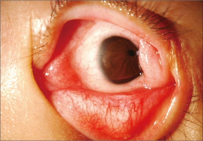
Anterior segment photographs showing lateral canthal deformity, symblepharon, and a conjunctival scar involving almost 360 degrees of peripheral cornea in the left eye.
An excisional biopsy, lateral canthoplasty, and symblepharon lysis with conjunctival autograft from the contralateral eye were performed in the left eye. Conjunctival scar tissue was excised and adhesion surrounding the lateral rectus muscle was completely relieved. To cover the bare sclera, a conjunctival graft was harvested from 3 clock hours of inferior bulbar conjunctiva of the contralateral eye. The conjunctival graft was sutured with surrounding conjunctiva using interrupted 7-0 Vicryl sutures. In addition, lateral canthoplasty using fornix forming sutures with 4-0 chromic catgut was also performed to reconstruct the lateral canthus.
Histopathologic examination revealed diffuse proliferation and infiltration of the collagenous tissue. These findings were consistent with a hypertrophic scar of conjunctival tissue (Fig. 2). Postoperative visual acuity was 20 / 100 OU. Exotropia was reduced to 20 prism diopters by the Krimsky method with full ductions and versions OS. There has been no evidence of recurrence over a 1-year follow-up period (Fig. 3).

Histopathologic examination reveals diffuse proliferation and infiltration of the collagenous tissue. These findings are consistent with hypertrophic scar. H&E, (A) ×100, (B) ×400.

(A) Preoperative photograph showing a temporal conjunctival scar with lateral canthal deformity of both eyes. (B,C) Photograph taken 1 year after surgery showing no recurrence and good cosmesis.
Discussion
ROP is characterized by retinal neovasculization affecting premature infants due to immature, incompletely vascularized retinas. Since the mid-1960s, neonatology practices have improved the survival rate of premature infants; this challenge urges the importance of management and treatment of ROP. From 1968, several reports have suggested cryotherapy of the peripheral retina for improvement of the course of the disease [1,2]. In order to resolve the uncertainty regarding the value of cryotherapy for ROP, a multicenter trial (the CRYO-ROP study) was designed to evaluate its safety and efficacy. A ten-year follow-up period to the CRYO-ROP study revealed that cryotherapy reduced unfavorable structural and functional outcomes by preventing progression of threshold disease [3].
Common complications related to cryotherapy include vitreous, retinal, and preretinal hemorrhage. Conjunctival chemosis, subconjunctival hemorrhage, conjunctival laceration, and hyphema have also been reported as anterior segment complications [1,4]. To the best of our knowledge, two reports on severe conjunctival lesions after treatment of ROP have been described in the literature. One report described a case of massive conjunctival proliferation following cryotherapy. In that report, the patient underwent repeated cryotherapy 8 times OD and 5 times OS for regression of extraretinal fibrovascular proliferation. No conjunctival peritomy was performed [5]. The other report described three cases of symblepharon after diode laser therapy. The authors suggested that symblepharon was related to a peritomy procedure during therapy, because no additional development of symblepharon occurred after cessation of the peritomy procedure [6]. Postoperative scarring and restriction can also develop after strabismus surgery. The scarring mainly involves muscle, Tenon's capsule, sclera and conjunctiva and it may arise from poor surgical technique or suture reaction. In this case, although the patient did not undergo repeated cryotherapy or conjunctival peritomy, a severe conjunctival hypertrophic scar and symblepharon developed as a complication after only one procedure of cryotherapy for ROP.
To avoid unexpected complications, surgeons who perform cryotherapy should exercise great skill and care. Moreover, this rare complication should be regarded as one of the differential diagnoses for an unusual conjunctival lesion in a patient with a history of ROP.
Notes
This paper was presented as a poster at the 103th annual meeting of the Korean Ophthalmological Society in April 2010.
No potential conflict of interest relevant to this article was reported.