


Dear Editor,
We would like to introduce a case of gouty tophus of eyelid, which is an unusual presentation of gout patients. Gouty tophus of eyelid is a rare condition; to our knowledge, no case of gouty tophus of eyelid has been reported in Korea.
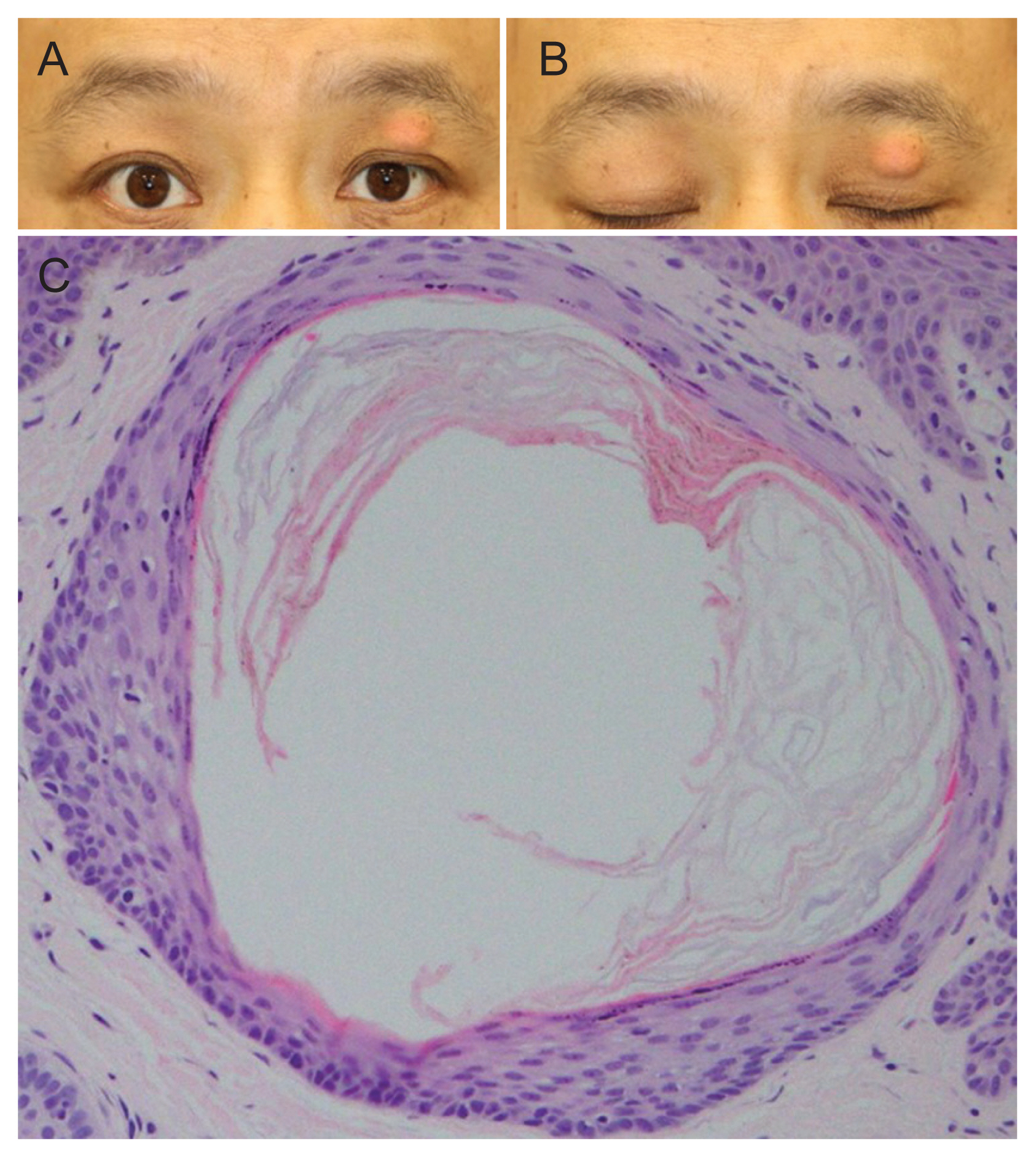
A 43-year-old male patient presented to us with left upper eyelid mass that gradually enlarged over 3 years. He had no other underlying diseases except gout and he was taking medication for gout. His uncorrected visual acuity was 20 / 20 in the right eye, 18 / 20 in the left eye. About 1.5 cm ├Ś 1.2 cm sized round, semi-soft, movable, subcutaneous yellowish mass with skin adhesion was visible at the left upper eyelid (Fig. 1A, 1B). He did not report any pain or tenderness around the mass lesion. Intraocular pressure was normal OU and anterior segment exam showed no abnormalities. We recommended him left upper eyelid mass excisional biopsy.
A mass was superficial to orbital septum. Mass was freed from adjacent tissues and removed en bloc for excisional biopsy.
The gross specimen was 1.5 cm ├Ś 0.7 cm ├Ś 0.7 cm sized round mass. Pathologic examination showed pools of deposition of amorphous material and its debris with foreign body reaction (Fig. 1C), consistent with gouty tophus.
Gout is an inflammatory arthritis which typically results from an increased uric acid level in our body [1]. Increased uric acid pool leads to collection of monosodium urate crystals in or around the joints, which is called tophi [2]. Tophus often affects peripheral joints such as joints of toe, metatarsophalangeal joints and knee, but many literatures report that tophus can build up at any part of our body [2]. Still, gouty tophus of the eyelid is an unusual manifestation of gout patients. To our knowledge, only 6 cases of gouty tophus in the periocular area were reported. Among six cases, two cases were medial canthal tophus, other two cases were lateral canthal tophus and the other two cases were eyelid tophus. Thus, this is the third report of gouty tophus of the eyelid as far as we know. Patients with gouty tophus of the eyelid often presents gradual enlargement of painless mass [3]. Definitive diagnosis of eyelid tophus is made by histopathologic findings [4]. Most important histopathologic finding of eyelid tophus is needle shaped monosodium urate crystals which are strongly negative birefringent under polarized light microscopy. The pathogenesis of tophus formation in the eye and the reason of its rarity are yet unknown [1].
In conclusion, we report a first Korean case of gouty tophus of the eyelid which is very unusual ocular manifestation of gout. Suspicion of eyelid tophus must be made in patients with painless eyelid mass who have history of gout. In order to confirm its diagnosis and exclude other types of masses, excisional biopsy and histopathologic review is mandatory [4].
 PDF Links
 PubReader
 ePub Link
 Full text via DOI
 Full text via PMC
 Download Citation
 Print
Print


