

Dear Editor,
Schwannoma is a benign peripheral nerve sheath tumor that frequently occurs in the orbit. However, an eyelid schwannoma is extremely rare [1], and only 20 cases have been reported. We report four cases of eyelid schwannoma and describe their common features.
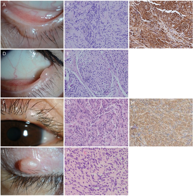
A 47-year-old man presented with a lower eyelid mass of the left eye for two years. The mass measured 6 mm ├Ś 3 mm, and it was not cystic, was non-pigmented, and without tenderness or erythema. The cilia on the surface of the mass were preserved (Fig. 1A). Shaving excisional biopsy revealed well-circumscribed elongated spindle cells forming palisades with strong S-100 positivity, consistent with eyelid schwannoma (Fig. 1B, 1C).
A 36-year-old woman was referred for evaluation of a lower eyelid mass of the left eye that had persisted for one year. Located on muco-cutaneous junction just above the cilia line, the mass measured 1.5 mm ├Ś 2.5 mm and was solid and amelanotic without inflammation (Fig. 1D). Shaving excisional biopsy demonstrated spindle cells forming a palisading pattern and a hyper/hypocellular area, consistent with eyelid schwannoma (Fig. 1E).
A 60-year-old woman presented with a lateral upper eyelid mass of the left eye. The 3 mm ├Ś 3 mm-sized mass was painless, non-pigmented and solid (Fig. 1F). A shaving excisional biopsy was performed, revealing spindle cells forming a palisading pattern and a Verocay body. Hyper/hypocellular area were also found with S-100 positivity, consistent with eyelid schwannoma (Fig. 1G, 1H). This patient has been previously reported [2].
A 45-year-old man was referred for a lateral upper eyelid mass of the right eye lasting for two years. A 5 mm ├Ś 3 mm-sized, solid, amelanotic mass without tenderness was observed (Fig. 1I). Shaving excisional biopsy revealed spindle cells with a palisading pattern and a hyper/hypocellular area, consistent with eyelid schwannoma (Fig. 1J).
We reported four cases of eyelid schwannoma, and each case demonstrated common features of this benign tumor: solid, non-pigmented mass with a smooth surface. Schwannoma similar to other benign eyelid tumors, such as the epidermal inclusion cyst, amelanotic nevus and chalazion [1]. However, the eyelid schwannoma differs from these conditions in that the epidermal inclusion cyst is cystic and often has a central pore [3] and the chalazion shows focal inflammatory signs [4].
The most distinguishing pathologic feature of schwannoma is reactivity to S-100, a protein only expressed in the central nervous system, Schwann cell and melanocyte [2].
The presence of multiple schwannomas is related to neurofibromatosis, although there is no relationship with neurofibromatosis for a solitary schwannoma [5]. All four cases in the present report had no relationship with neurofibromatosis. Although malignant transformation has not been reported, schwannomas should be biopsied and completely excised with a negative margin to prevent recurrence with more aggressive behaviors.
In conclusion, we reported four cases of eyelid schwannoma. Eyelid schwannoma should be considered as one of the differential diagnoses when a non-pigmented solid eyelid mass with a smooth surface is encountered.
 PDF Links
 PubReader
 Full text via DOI
 Full text via PMC
 Download Citation
 Print
Print


