

Cancer stem cells (CSCs) have been introduced and investigated as a new hypothesis of tumorigenesis in recent years [1, 2]. In this theory, a population of cells with stem cell- like characteristics, i.e. self-renewal, multipotency, and tumor initiation capability, exists in tumors. This population gives rise to the bulk of the tumor cells with more differentiated phenotypes. CSCs could be helpful not only in understanding the pathogenesis of the cancer, but also in the treatment of the cancer itself. CSCs were regarded as the key component of chemoresistance of cancers, and it should be the target for drug development. Recent study revealed that selective targeting of CSCs is possible [3].
CSCs, like normal stem cells, are believed to exist within a protective perivascular niche, but can escape from the niche control. The presence of a perivascular niche was proved in the hematopoietic system, as well as in brain tumors [4, 5]. Calabrese et al. [5] suggest that CSCs in brain tumors, similar to neural stem cells, reside in vascular niches that are important targets for therapeutic approaches. A few orthotopic xenograft studies in brain tumors suggest that CSCs secrete angiogenic factors, such as vascular endothelial growth factor (VEGF), that promote the recruitment and formation of tumor blood vessels [5, 6].
Retinoblastoma is the most common intraocular childhood tumor [7]. Moreover, retinoblastoma is believed to be a good model in which to search for a tumor cell of origin and to understand the molecular pathogenesis of malignant tumors [8, 9]. Recent studies demonstrate that a small proportion of human and mouse retinoblastoma cell populations express cancer stem cell markers (including ABCG2) and neural stem cell markers (including MCM2) [10, 11]. Zhong et al. [12] demonstrated the presence of tumorigenic retinal stem-like cells that contribute to tumorigenesis in human retinoblastoma.
Without regard to relation with cancer stem cells or a perivascular niche, VEGF has been studied as an important angiogenic growth factor in retinoblastoma [13, 14]. In recent studies, anti-angiogenic therapy targeting VEGF has been proposed as a promising new treatment strategy of retinoblastoma [15-17].
In this study, we investigated the expression of ABCG2 and MCM2 proteins in human retinoblastoma tissues and the correlation of their expression with VEGF proteins, which may be related to the CSCs-perivascular niche complex. The purpose of this study was to evaluate the relation between VEGF and the cancer stem cell-vascular niche complex in human retinoblastoma tissue.
Materials and Methods
Patients
Six patients who were diagnosed as having unilateral highly progressive large retinoblastoma, group 5a in Reese-Ellsworth classification, were included in this study. All patients underwent enucleation without chemotherapy or focal therapies. All human retinoblastoma tissue samples were obtained with informed consent and in accordance with the tenets of the Declaration of Helsinki.
Antibodies and chemicals
For the detection of ABCG2 protein, mouse monoclonal anti-ABCG2 antibody (1:100, Santa Cruz Biotechnology, Santa Cruz, CA, USA) was used. Primary goat polyclonal anti-MCM2 antibody (1:200, Santa Cruz Biotechnology) and primary rabbit polyclonal anti-VEGF antibody (1:100, Santa Cruz Biotechnology) were also used for staining.
Immunofluorescence
Paraffin-embedded enucleated eyes were sectioned into 4-µm sections. Sections of archival human specimens were rehydrated with xylene and graded alcohols. After washing in running water for five minutes, antigen retrieval was performed by placing slides in 10 mM sodium citrate buffer (pH 6.0), heated at 120℃ for 10 minutes. The slides and buffer were then cooled to room temperature for 30 minutes. After washing with phosphate buffered saline (PBS), the slides were incubated with 0.2% Tripton X-100 for 15 minutes. After three rinses in PBS for five minutes each, sections were incubated with blocking solution for 30 minutes, followed by incubation with primary antibodies: five hours for ABCG2 and VEGF and two hours for MCM2. All sections were incubated for two hours with secondary antibody, followed by mounting.
Results
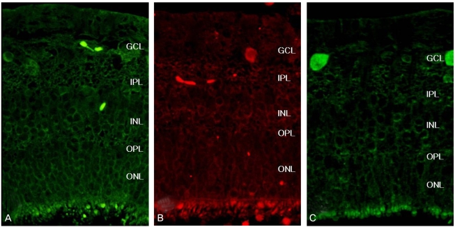
ABCG2, MCM2 and VEGF expression in the non-neoplastic retina
ABCG2 and MCM2 were sporadically expressed in the ganglion cell layer, the inner nuclear layer, and the photoreceptor layer in the non-neoplastic portion of the retina of the tumor-bearing eye. Expression of these markers seemed not to be related with retinal vasculature (Fig. 1A and 1B).
VEGF was sporadically expressed in the inner retina where retinal vessels are abundantly distributed (Fig. 1C).
ABCG2, MCM2 and VEGF expression in retinoblastoma tumors
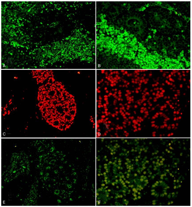
ABCG2 was strongly expressed in the Wintersteiner rosettes or tumor lobules. In the rosettes or lobules, sporadic expression of ABCG2 was observed and not related to tumor vasculature. Most of ABCG2-stained cells were adjacent to the rosettes (Fig. 2A and 2B).
MCM2 was strongly positive in the rosettes, especially in the outer portion of the rosettes (Fig. 2C and 2D). Within the lobules of the tumor, MCM2 was expressed around the blood vessels.
VEGF was expressed in the rosettes or tumor lobules, similar to MCM2. But unlike MCM2, VEGF was strongly expressed in the inner portion of the rosettes (Fig. 2E and 2F). In the lobules of retinoblastoma, VEGF was also sporadically expressed, but not directly associated with tumor vessels.
Discussion
Evidence suggests that CSCs exist in human retinoblastoma [10, 11, 18]. Seigel et al. [18] observed the presence of a small population which express a cancer stem cell marker (ABCG2) and a neural stem cell marker (MCM2) in tumors from transgenic mice, human retinoblastoma cell lines, and human retinoblastomas. ABCG2 is a half ATP-binding cassette transporter in the G2 subfamily that has been utilized to identify stem cells in several types of cancer. ABCG2 is also related to a resistance to a wide variety of anticancer drugs [19]. MCM2 is one of six members of the family of minichromosome maintenance proteins involved in initiating DNA replication [20]. MCM2 is a proven marker of neural stem cells and is known to be related to the aggressiveness of neuroectodermal tumors and retinoblastoma [11, 21].
In this study, the expression of ABCG2 and MCM2 in human archival retinoblastoma tumors and the non-neoplastic retina in the same eye was observed by immunofluorescence staining. Concurrent with previous studies, MCM2 positivity was observed surrounding blood vessels in both well- and poorly-differentiated tumor tissues. MCM2 was strongly positive in tumor rosettes. In normal retinas, MCM2 positivity was observed in the ganglion cell layer, inner nuclear layer, and photoreceptor layer. The ABCG2 staining pattern was similar in the normal retina. In retinoblastoma tumor tissues, on the other hand, ABCG2 positivity was observed outside of the tumor rosettes and lobules. This result differed from previous reports. With our data, ABCG2 and MCM2 stained different portions of the retinoblastoma tumor tissue.
According to the cancer stem cell hypothesis, the cancer stem cell-vascular niche is an important aspect in understanding CSCs and in differentiating them from normal stem cells. Stem cells reside in "niches", defined as physical spaces in the tissue used to maintain homeostasis [4]. CSCs might escape from normal niche control, and the dysregulated stem cell division may give rise to turmor- igenesis. CSCs secrete angiogenic factors, such as VEGF, that promote the recruitment and formation of tumor vasculature [5, 6].
VEGF had been studied as an important regulator of physiologic and pathologic angiogenesis before the concept of CSCs emerged. VEGF expression in retinoblastoma has also been studied because retinoblastomas are highly vascularized tumors that are dependent on angiogenesis [13, 14, 22]. Recent studies have revealed that VEGF-targeted treatments suppress tumor angiogenesis and growth significantly in retinoblastomas. In these studies, the anti- angiogenic effect by blocking VEGF was considered the most important mode of action [15-17]. Until now, the mechanism of action with anti-VEGF agents has not been fully understood [23].
In this study, we speculate that VEGF is related to CSCs or the CSCs-perivascular niche complex. VEGF is known to be secreted from neoplastic cells and influences endothelial cells as paracrine mediators, but the relationship between VEGF and CSCs in retinoblastomas is unknown. We stained the VEGF protein in retinoblastoma tissues to locate the expression and to compare it to that of ABCG2 and MCM2. Consistent with previous studies, VEGF immunoreactivity was observed sporadically at the inner retina where there is abundant retinal vasculature in the normal retina. However, this staining was weak and not directly related to retinal vasculature. In retinoblastoma tumor tissues, VEGF expression was observed in the rosettes or tumor lobules, similar to MCM2 expression. Unlike MCM2, strong VEGF expressed was observed in the inner portion of the rosettes. We have no immediate explanation for the difference in the distribution of these proteins. One possible reason that VEGF expression is observed at the inner portion of the rosettes is that it is a secretary protein. The lobules of the retinoblastoma also showed sporadic VEGF positivity but that was not directly associated with tumor vessels. If CSCs or the CSCs-perivascular niche have pivotal roles in VEGF secretion, VEGF immunopositivity must be observed in similar locations as CSC markers. Similarly, if VEGF directly contributes to the CSCs-perivascular niche complex, then VEGF must be located around the tumor vasculature. However, our data show that VEGF expression is not related to ABCG2 expression or tumor vasculature. Instead, VEGF expression was similar to that of MCM2 except with specific topographical distribution. Our study suggests that, although VEGF might not directly contribute to the CSC-perivascular niche complex, it could play some role in the differentiation of tumor cells and in rosette formation.
 PDF Links
 PubReader
 Full text via DOI
 Full text via PMC
 Download Citation
 Print
Print


